Greetings Card > Arts > Artists > G > Thomas Gray
Greetings Card : Purkinje nerve cells in the cerebellum


![]()
Cards from Science Photo Library
Purkinje nerve cells in the cerebellum
Purkinje cells in the cerebellum. Fluorescent light micrograph of Purkinje cells (green) in the cerebellum of the brain. Purkinje nerve cells have a flask-like body from which numerous highly branched dendrites extend. They are found in the grey matter (cortex) of the cerebellum, at the boundary between the granular layer (blue/red) and the molecular layer (red/green). The dendrites relay signals to the cell body, which passes them on through its single axon (green) in the granular layer. The cerebellum is a structure at the base of the brain that plays an important role in motor control, sensory perception and learning. Magnification: x200 when printed 10cm wide
Science Photo Library features Science and Medical images including photos and illustrations
Media ID 6449277
© THOMAS DEERINCK, NCMIR/SCIENCE PHOTO LIBRARY
Cerebellar Cerebellum Cortex Dendrite Dendrites Fluorescent Light Micrograph Granular Layer Gray Grey Matter Histological Histology Molecular Layer Nerve Cell Neuron Purkinje Cell Brain Light Microscope Nervous System Neurological Neurology
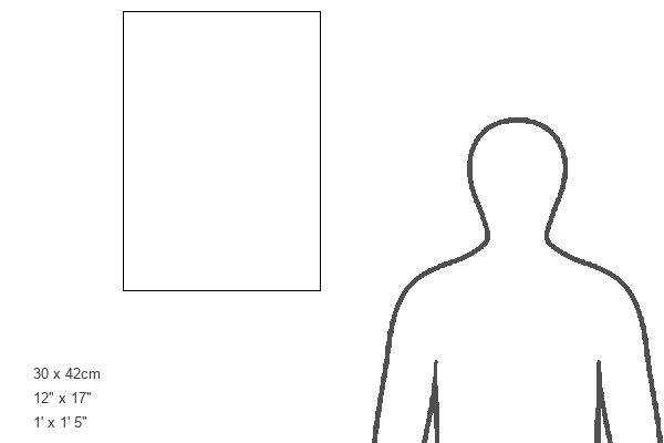
Greetings Card Large (A4)
Discover the beauty of neuroscience with our unique range of Science Greetings Cards from Media Storehouse. Featuring an captivating image of Purkinje nerve cells in the cerebellum, these cards showcase the intricate details of these vital brain cells. Illuminated by fluorescent light, the green Purkinje cells stand out against the backdrop of the cerebellum, their distinctive flask-like bodies and extensive dendrites a testament to the complex network of the human nervous system. Perfect for the science enthusiast or anyone who appreciates the wonders of the natural world, these cards are a thoughtful and engaging way to express your sentiments. Order yours today and brighten someone's day with a touch of scientific wonder.
Create your own large greetings card. Size when folded is A4 (21x30cm or 8.3x11.7 inches)
Greetings Cards suitable for Birthdays, Weddings, Anniversaries, Graduations, Thank You and much more
Estimated Image Size (if not cropped) is 29.7cm x 21cm (11.7" x 8.3")
Estimated Product Size is 29.7cm x 42cm (11.7" x 16.5")
These are individually made so all sizes are approximate
Artwork printed orientated as per the preview above, with landscape (horizontal) orientation to match the source image.
FEATURES IN THESE COLLECTIONS
> Animals
> Mammals
> Muridae
> Blue-grey Mouse
> Arts
> Artists
> G
> Thomas Gray
> Science Photo Library
> Specialist Imaging
EDITORS COMMENTS
This image showcases a fluorescent light micrograph of Purkinje cells in the cerebellum of the human brain. Purkinje cells, named after the Czech anatomist Jan Evangelista Purkinje who first described them in 1837, are large, flask-shaped nerve cells located in the grey matter (cortex) of the cerebellum. They are easily distinguishable due to their unique morphology, characterized by a large, flask-like soma from which numerous highly branched dendrites extend. The dendrites of Purkinje cells are the primary site of reception and integration of signals from the granular layer below, which are then relayed to the cell body. The cell body passes these signals on through its single axon, which extends into the granular layer and forms synapses with the dendrites of granule cells. The cerebellum, a structure located at the base of the brain, plays a crucial role in motor control, sensory perception, and learning. In this image, the Purkinje cells are situated at the boundary between the granular layer (stained blue/red) and the molecular layer (stained red/green). The molecular layer is where the Purkinje cell axons form synapses with mossy fibers, initiating the complex neural networks that underpin the functions of the cerebellum. Magnified at 200x when printed 10cm wide, this mesmerizing snapshot offers a glimpse into the intricate world of the human nervous system. The delicate balance and intricate connections between these cells underscore the complexity and sophistication of the brain's neural architecture.
MADE IN THE UK
Safe Shipping with 30 Day Money Back Guarantee
FREE PERSONALISATION*
We are proud to offer a range of customisation features including Personalised Captions, Color Filters and Picture Zoom Tools
SECURE PAYMENTS
We happily accept a wide range of payment options so you can pay for the things you need in the way that is most convenient for you
* Options may vary by product and licensing agreement. Zoomed Pictures can be adjusted in the Basket.