
Purkinje Cell Collection
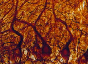
The intricate world of the cerebellum tissue comes to life in this captivating light micrograph


All Professionally Made to Order for Quick Shipping
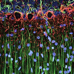
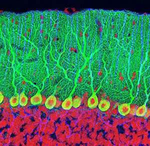
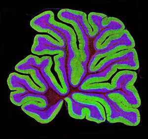
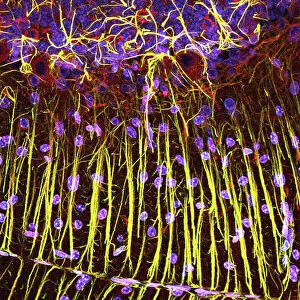
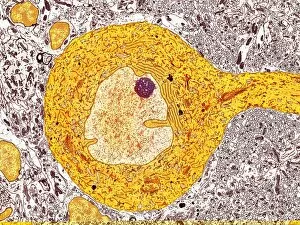
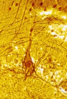
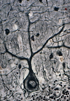
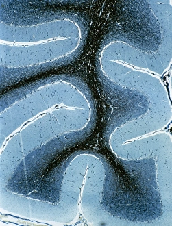
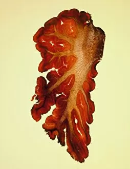
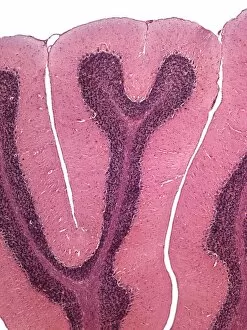
The intricate world of the cerebellum tissue comes to life in this captivating light micrograph. Amongst its fascinating structures, we find the Purkinje nerve cells standing out like stars in a dark sky. These specialized neurons play a crucial role in coordinating movement and maintaining balance. Zooming closer into the cerebellum structure, we witness the elegance of these Purkinje nerve cells once again through another mesmerizing light micrograph. Their distinct shape and branching dendrites create a unique pattern that sets them apart from other neurons. Intriguingly, an even more detailed view is unveiled through TEM C014 / 0583, revealing the inner workings of a single Purkinje nerve cell. This high-resolution image showcases their elongated body with numerous dendritic spines reaching out like delicate branches seeking connections with other neurons. Returning to the beauty of cerebellum tissue under different angles, multiple light micrographs provide us with an array of perspectives on these remarkable cells. Each image captures their presence amidst a sea of interconnected neural networks within this vital brain region. As we delve deeper into understanding our complex nervous system, it is awe-inspiring to witness such intricacy at work within every corner of our brains. The Purkinje nerve cells stand as testament to nature's ingenuity and remind us that there are still countless wonders waiting to be discovered within ourselves.