
Dendrite Collection
"Dendrite: The Intricate Network of Communication in the Cerebellum" In the vast expanse of our brain, lies a fascinating structure known as the cerebellum


All Professionally Made to Order for Quick Shipping
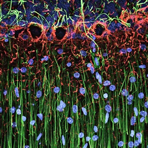
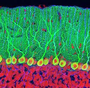
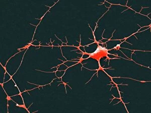
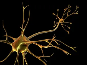
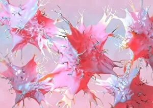
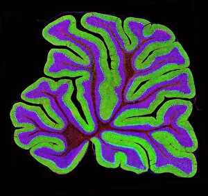
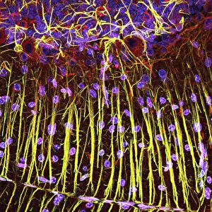
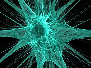
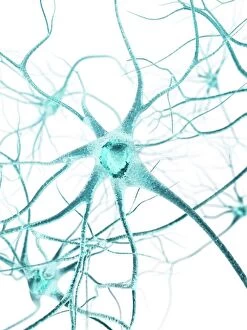
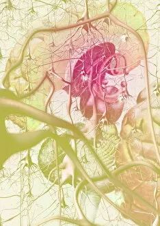
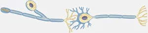
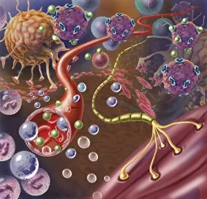
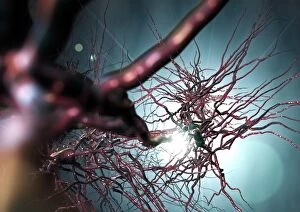
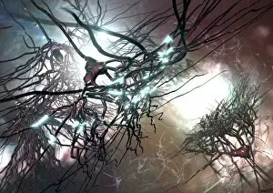
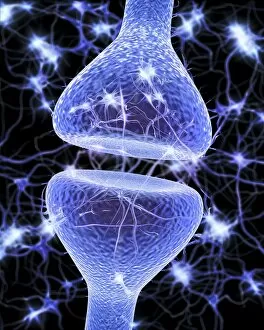
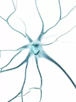
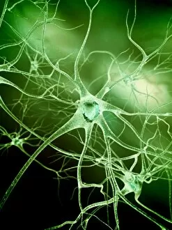
"Dendrite: The Intricate Network of Communication in the Cerebellum" In the vast expanse of our brain, lies a fascinating structure known as the cerebellum. Within this intricate tissue, light micrographs reveal a mesmerizing world of dendrites - the key players in nerve cell communication. Underneath the lens, we witness the beauty and complexity of purkinje nerve cells in the cerebellum. These remarkable structures extend their branches like delicate trees, reaching out to connect with other neurons through their dendritic extensions. Through scanning electron microscopy (SEM), we delve deeper into this realm of neural connectivity. Nerve cells come alive before our eyes, showcasing their unique shapes and patterns that form an elaborate network within our brains. Artwork depicting dendritic cells captivates us with its abstract representation. It serves as a reminder that these microscopic entities hold immense power in transmitting electrical signals across synapses – enabling us to move, think, and feel. The cerebellum's structure is unveiled once again through light micrography - revealing its densely packed motor neurons intricately intertwined with countless dendrites. This organized chaos forms the foundation for precise coordination and control over our movements. As we explore further into this captivating world, more light micrographs showcase cerebellar tissues teeming with life. Each image tells a story of interconnectedness between nerve cells - an orchestra playing harmoniously to ensure smooth functioning of our body and mind. From scientific imagery to artistic interpretations, dendrites continue to fascinate us by bridging gaps between neurons both physically and metaphorically, and are not mere extensions but rather conduits for information flow within our complex neural circuitry. So let us marvel at these incredible structures that shape who we are - reminding ourselves that beneath every thought or action lies an intricate web woven by dendrites; connecting us all on a fundamental level within the labyrinthine landscape of our minds.