Fraud Blocker Skip to main content

Cortex Collection

The cortex, a fascinating part of the brain, is responsible for numerous functions that are essential to our daily lives

Background imageCortex Collection: Purkinje nerve cells in the cerebellum

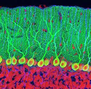
Purkinje nerve cells in the cerebellum
Purkinje cells in the cerebellum. Fluorescent light micrograph of Purkinje cells (green) in the cerebellum of the brain. Purkinje nerve cells have a flask-like body from which numerous highly

Background imageCortex Collection: Motor homunculus

Motor homunculus
This model shows what a mans body would look like if each part grew in proportion to the area of the cortex of the brain concerned with its movement

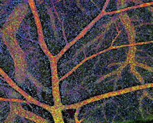
Background imageCortex Collection: Brain tissue blood supply

Brain tissue blood supply. Light micrograph of a section through cortex tissue from a brain, showing the blood vessels (branching) that supply it

Background imageCortex Collection: Vascular bundle, SEM

Vascular bundle, SEM
Vascular bundle. Coloured scanning electron micrograph (SEM) of a section through a vascular bundle from a rootlet of a dicotyledon plant

Background imageCortex Collection: Basal ganglia, artwork

Basal ganglia, artwork
Basal ganglia. Computer artwork of the human brain, showing some of the structures that make up the basal ganglia (basal nuclei, pink)

Background imageCortex Collection: Purkinje nerve cells in the cerebellum

Purkinje nerve cells in the cerebellum
Purkinje cells in the cerebellum. Fluorescent light micrograph of Purkinje cells (green) in the cerebellum of the brain. Purkinje nerve cells have a flask-like body from which numerous highly

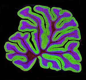
Background imageCortex Collection: Cerebellum structure, light micrograph

Cerebellum structure, light micrograph
Cerebellum structure. Fluorescent light micrograph of a section through the cerebellum of the brain. The cerebellum comprises three main layers

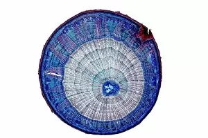
Background imageCortex Collection: Lime tree stem, light micrograph

Lime tree stem, light micrograph
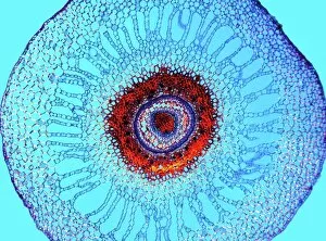
Lime tree stem. Light micrograph of a section through the stem of a lime tree (Tilia europaea). The outer epidermis has been shed and replaced by a layer of cork (dark red)

Background imageCortex Collection: Phantom pain after amputation, artwork

Phantom pain after amputation, artwork
Phantom pain after amputation. Artwork of a man experiencing phantom pain (red flash) from his amputated hand (left). This is caused by stimuli to the motor and pre-motor cortical areas of his brain

Background imageCortex Collection: False-colour SEM showing two renal glomeruli

False-colour SEM showing two renal glomeruli
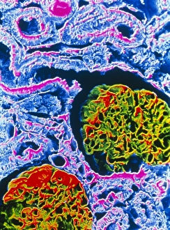
False-colour scanning electron micrograph of a section of kidney cortex, showing two glomeruli (red & green). A glomerulus is a tiny knot of blood capillaries which lies cupped within a Bowmans

Background imageCortex Collection: Water fern rhizome, light micrograph

Water fern rhizome, light micrograph
Water fern rhizome. Polarised light micrograph of a section through a rhizome from a water fern (Marselia vestita). Under the epidermis (the outer layer)

Background imageCortex Collection: Pine tree stem, light micrograph

Pine tree stem, light micrograph
Pine tree stem. Coloured light micrograph of a cross-section through the stem of a pine tree. (Pinus sp.). The red wood (xylem, lower left) has resin canals in it, which are outlined in green

Background imageCortex Collection: Maiden hair tree stem, SEM

Maiden hair tree stem, SEM
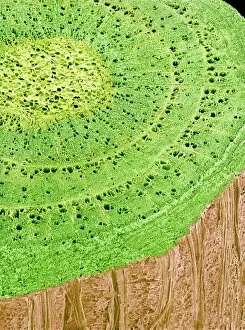
Maiden hair tree stem. Coloured scanning electron micrograph (SEM) of a section through a maidenhair tree (Ginkgo biloba) stem. At centre is the pith, which consists of soft, spongy parenchyma cells

Background imageCortex Collection: Anatomy of adrenal gland, cross section

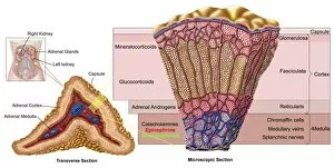
Anatomy of adrenal gland, cross section

Background imageCortex Collection: Purple flowered red Peruvian bark tree, Cinchona cortex peruvianus ruber

Purple flowered red Peruvian bark tree, Cinchona cortex peruvianus ruber
5856673 Purple flowered red Peruvian bark tree, Cinchona cortex peruvianus ruber by Sowerby, James (1757-1822); (add.info.: Purple flowered red Peruvian bark tree, Cinchona cortex peruvianus ruber)

Background imageCortex Collection: Gray matter of the cerebral cortex, Meynert (engraving)

Gray matter of the cerebral cortex, Meynert (engraving)
3634183 Gray matter of the cerebral cortex, Meynert (engraving) by English School, (19th century); Private Collection; (add.info.: Gray matter of the cerebral cortex, Meynert)

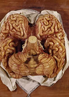
Background imageCortex Collection: Model of a brain (wax)

Model of a brain (wax)
BEN360191 Model of a brain (wax) by Susini, Clemente (1754-1814); Museo della Specola, Florence, Italy; (add.info.: cervello; scultura di cera; ); Photo eRaffaello Bencini; Italian, out of copyright

Background imageCortex Collection: Brain blood vessels, artwork

Brain blood vessels, artwork
Brain blood vessels. Computer artwork showing the blood vessels that supply the human brain. The front of the brain is at left

Background imageCortex Collection: Anatomy of human head with skull and brain superimposed

Anatomy of human head with skull and brain superimposed, side view

Background imageCortex Collection: Thymus gland, light micrograph C015 / 4970

Thymus gland, light micrograph C015 / 4970
Thymus gland. Light micrograph of a section through tissue from the thymus gland, part of the lymphoid system. The dense areas are the thymic cortex, which produces lots of lymphocytes

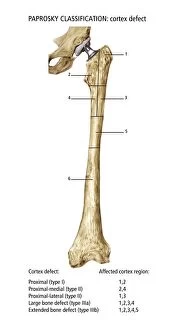
Background imageCortex Collection: Paprosky femur defect classification C016 / 6621

Paprosky femur defect classification C016 / 6621
Paprosky femur defect classification. Diagram showing the classification system for femur cortex defects used to assess revision (replacement or repair) of a hip implant

Background imageCortex Collection: Kidney anatomy, artwork

Kidney anatomy, artwork
Kidney anatomy. Cutaway artwork showing the internal structure of a kidney. The kidney, one of a pair of such organs, is part of the human excretory system

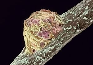
Background imageCortex Collection: Root nodule

Root nodule. Coloured scanning electron micrograph (SEM) of a root nodule on a pea plant (Pisum sativum) caused by the nitrogen-fixing soil bacteria Rhizobium leguminosarum

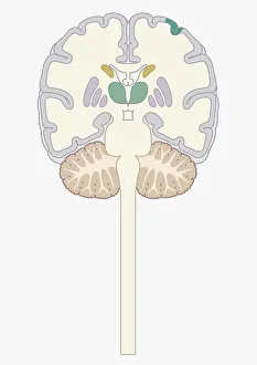
Background imageCortex Collection: Digital cross section illustration of human brain

Digital cross section illustration of human brain

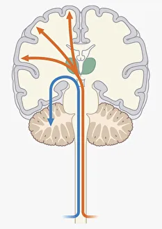
Background imageCortex Collection: Digital illustration of pain signal to area of somatosensory cortex in human brain via spine

Digital illustration of pain signal to area of somatosensory cortex in human brain via spine

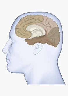
Background imageCortex Collection: Digital illustration of head in profile showing medial view of cortex in human brain

Digital illustration of head in profile showing medial view of cortex in human brain

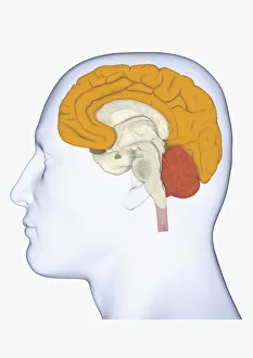
Background imageCortex Collection: Digital illustration head in profile with cerebrum and cortex highlighted

Digital illustration head in profile with cerebrum and cortex highlighted

Background imageCortex Collection: Digital illustration of head in profile showing lateral view of cortex in brain

Digital illustration of head in profile showing lateral view of cortex in brain

Background imageCortex Collection: Digital illustration of various areas of cortex in human brain receiving input from sense organs

Digital illustration of various areas of cortex in human brain receiving input from sense organs

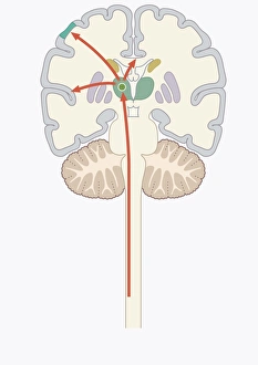
Background imageCortex Collection: Digital illustration of unconscious and conscious pathways in human brain passing through thalamus

Digital illustration of unconscious and conscious pathways in human brain passing through thalamus ending in parietal lobe of cortex

Background imageCortex Collection: Anatomy of human bone marrow

Anatomy of human bone marrow

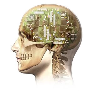
Background imageCortex Collection: Male human head with skull and artificial electronic circuit brain

Male human head with skull and artificial electronic circuit brain in ghost effect, side view

Background imageCortex Collection: Cutaway view of the human kidney and adrenal gland

Cutaway view of the human kidney and adrenal gland

Background imageCortex Collection: Ewings sarcoma locations on the skeleton

Ewings sarcoma locations on the skeleton and detail of tumor on head of femur

Background imageCortex Collection: Prion disease treatment, artwork C018 / 1838

Prion disease treatment, artwork C018 / 1838
Prion disease treatment. Artwork of three pyramidal neurons (nerve cells). The cell at left is healthy. The cell at back has died because of a prion (pink) infection

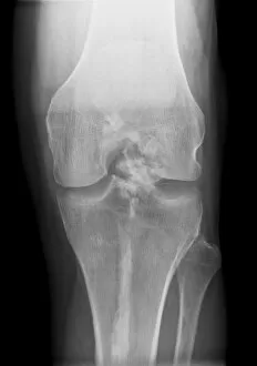
Background imageCortex Collection: Melorheostosis of the knee, X-ray C017 / 7146

Melorheostosis of the knee, X-ray C017 / 7146
Melorheostosis of the knee. X-ray of left knee of a 52 year old male patient showing damage (white area at centre) caused by melorheostosis

Background imageCortex Collection: Human brain, artwork F006 / 6313

Human brain, artwork F006 / 6313
Human brain, computer artwork. The front of the brain is at bottom

Background imageCortex Collection: Nerve cells, artwork F007 / 5514

Nerve cells, artwork F007 / 5514
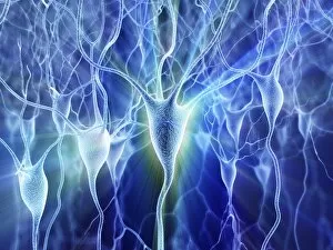
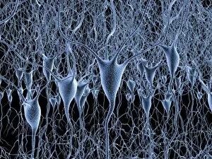
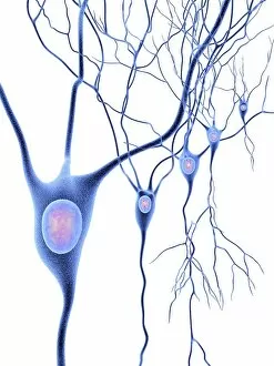
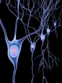
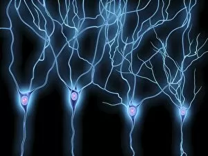
Computer artwork of nerve cells, also called neurons. Neurons are responsible for passing information around the central nervous system (CNS) and from the CNS to the rest of the body

Background imageCortex Collection: Nerve cells, artwork F007 / 5522

Nerve cells, artwork F007 / 5522
Computer artwork of nerve cells, also called neurons. Neurons are responsible for passing information around the central nervous system (CNS) and from the CNS to the rest of the body

Background imageCortex Collection: Nerve cells, artwork F007 / 5523

Nerve cells, artwork F007 / 5523
Computer artwork of nerve cells, also called neurons. Neurons are responsible for passing information around the central nervous system (CNS) and from the CNS to the rest of the body

Background imageCortex Collection: Nerve cells, artwork F007 / 5526

Nerve cells, artwork F007 / 5526
Computer artwork of nerve cells, also called neurons. Neurons are responsible for passing information around the central nervous system (CNS) and from the CNS to the rest of the body

Background imageCortex Collection: Nerve cells, artwork F007 / 5521

Nerve cells, artwork F007 / 5521
Computer artwork of nerve cells, also called neurons. Neurons are responsible for passing information around the central nervous system (CNS) and from the CNS to the rest of the body

Background imageCortex Collection: Nerve cells, artwork F007 / 5512

Nerve cells, artwork F007 / 5512
Computer artwork of nerve cells, also called neurons. Neurons are responsible for passing information around the central nervous system (CNS) and from the CNS to the rest of the body

Background imageCortex Collection: Nerve cells, artwork F007 / 5510

Nerve cells, artwork F007 / 5510
Computer artwork of nerve cells, also called neurons. Neurons are responsible for passing information around the central nervous system (CNS) and from the CNS to the rest of the body

Background imageCortex Collection: Nerve cells, artwork F007 / 5530

Nerve cells, artwork F007 / 5530
Computer artwork of nerve cells, also called neurons. Neurons are responsible for passing information around the central nervous system (CNS) and from the CNS to the rest of the body

Background imageCortex Collection: Nerve cells, artwork F007 / 5519

Nerve cells, artwork F007 / 5519
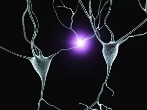
Computer artwork of firing nerve cells, also called neurons. Neurons are responsible for passing information around the central nervous system (CNS) and from the CNS to the rest of the body

Background imageCortex Collection: Nerve cells, artwork F007 / 5518

Nerve cells, artwork F007 / 5518
Computer artwork of nerve cells, also called neurons. Neurons are responsible for passing information around the central nervous system (CNS) and from the CNS to the rest of the body

Background imageCortex Collection: Nerve cells, artwork F007 / 5513

Nerve cells, artwork F007 / 5513
Computer artwork of nerve cells, also called neurons. Neurons are responsible for passing information around the central nervous system (CNS) and from the CNS to the rest of the body


All Professionally Made to Order for Quick Shipping

The cortex, a fascinating part of the brain, is responsible for numerous functions that are essential to our daily lives. One remarkable aspect of the the motor homunculus, a representation of how different parts of our body are controlled by specific areas in the brain. This intricate map showcases the complexity and precision with which movements are coordinated. Purkinje nerve cells in the cerebellum play a crucial role in maintaining balance and coordination. These unique cells have an elaborate branching structure that allows them to transmit information efficiently within this important region of the brain. Examining a vascular bundle under scanning electron microscopy reveals its intricate network, highlighting how blood vessels supply vital nutrients and oxygen to sustain healthy brain tissue. Artwork depicting basal ganglia captures their significance as they contribute to various functions such as movement regulation and decision-making processes. Their interconnectedness emphasizes their influence on overall brain function. Understanding the complex system that ensures proper blood supply to brain tissue is crucial. The delicate balance between oxygen delivery and demand plays a critical role in maintaining optimal cognitive function. A light micrograph showcasing a lime tree stem provides insight into plant anatomy but also serves as a reminder of nature's intricate design parallels with human biology. Similar Purkinje nerve cells can be found within the cerebellum structure when observed under light microscopy. Their distinct appearance further emphasizes their importance in coordinating movements smoothly and precisely. Phantom pain after amputation remains an enigma; artwork capturing this phenomenon highlights its impact on individuals who experience sensations from missing limbs—an intriguing area where neuroscience continues to seek answers. Exploring water fern rhizome under light microscopy offers glimpses into its internal structures, reminding us that biological intricacies extend beyond humans alone—a testament to nature's diversity and complexity. A pine tree stem captured through light microscopy unveils its cellular composition—underscoring similarities between plant tissues and those found within our own bodies—a reflection of shared evolutionary history across species boundaries.