
Grey Matter Collection
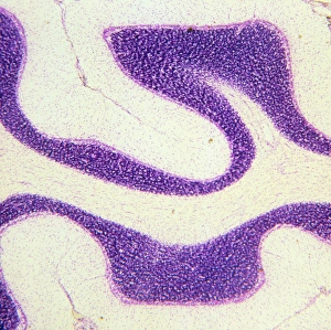
"Exploring the Intricacies of Grey Matter: A Journey into the Depths of Cerebellum Tissue" In this captivating light micrograph


All Professionally Made to Order for Quick Shipping
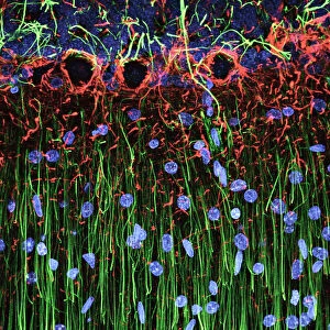
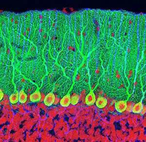
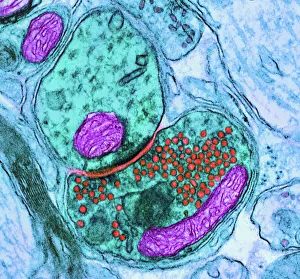
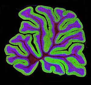
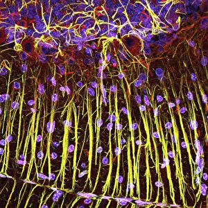
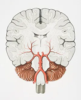
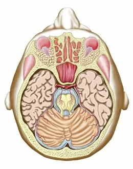
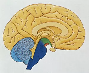
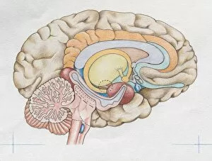
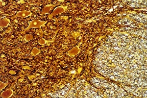
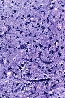
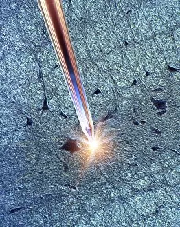
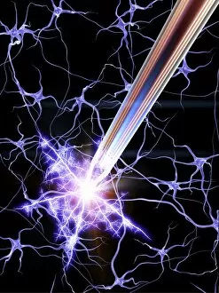
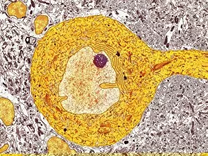
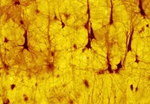
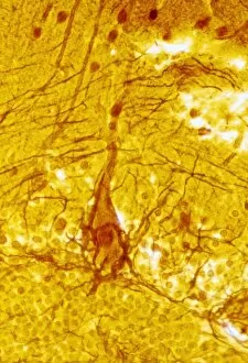
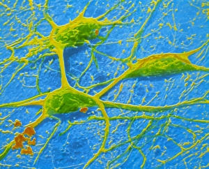
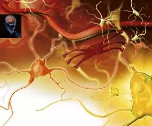
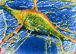
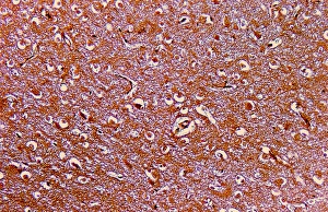
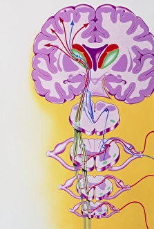
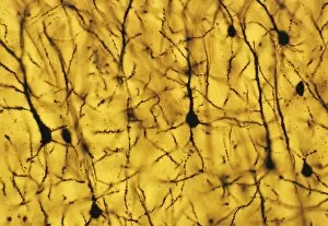
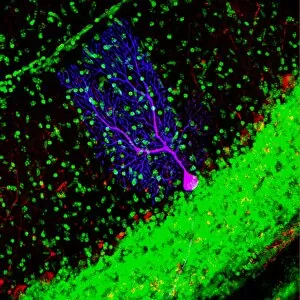
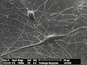
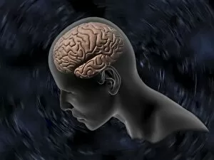
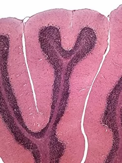
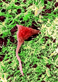
"Exploring the Intricacies of Grey Matter: A Journey into the Depths of Cerebellum Tissue" In this captivating light micrograph, we delve into the enigmatic world of grey matter, specifically focusing on the cerebellum tissue. The intricate network of synapse nerve junctions can be observed in a striking TEM image, showcasing their vital role in transmitting information within our brain. One cannot help but marvel at the elegance and complexity of Purkinje nerve cells found within the cerebellum. These remarkable cells are captured beautifully in both light micrographs and TEM images, highlighting their unique structure and function. Their significance lies in regulating motor coordination and balance, making them indispensable for our daily movements. Zooming out to view a broader perspective, a mesmerizing light micrograph reveals the overall structure of the cerebellum. Its distinct layers and organization come to life as we witness its crucial role in fine-tuning our motor skills. Further exploration takes us beyond just one region as we encounter various sections of the brain. A coronal cross-section offers an intriguing glimpse into its multidimensional nature while a transverse section provides insight into specific areas like midbrain. Finally, we arrive at perhaps one of nature's most fascinating creations -the human brain itself. Through a basic cutaway illustration, we witness key components such as thalamus, cerebrum, hypothalamus intricately intertwined with grey matter. This serves as a reminder that grey matter is not merely an abstract concept but rather an essential part that orchestrates our thoughts, emotions, memories- shaping who we are as individuals. As we embark on this journey through microscopic wonders and macroscopic complexities alike; let us appreciate how grey matter holds immense power within its delicate confines- connecting every aspect of our being with astonishing precision.