Greetings Card : Hartmannella vermiformis protozoa cysts C016 / 9402


![]()
Cards from Science Photo Library
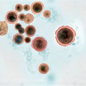
Hartmannella vermiformis protozoa cysts C016 / 9402
Hartmannella vermiformis protozoa cysts. Transmission electron micrograph (TEM) of a section through cysts (round) containing Hartmannella vermiformis protozoa. H. vermiformis is a single- celled organism that is found in soil and freshwater and feeds on bacteria
Science Photo Library features Science and Medical images including photos and illustrations
Media ID 9244053
© AMI IMAGES/SCIENCE PHOTO LIBRARY
Amoeboid Cyst Cysts Electron Microscope Histological Histology Infected Infecting Infection Micro Organism Micro Organisms Microbiology Microorganism Microorganisms Parasite Parasites Parasitic Protozoa Protozoan Protozoans Single Celled Tissue Transmission Electron Micrograph Unicellular Abnormal Amoebozoa Microbiological Section Sectioned Unhealthy
Greetings Card Large (A4)
Discover the intriguing world of microscopic organisms with our Hartmannella vermiformis Protozoa Cysts Greetings Cards from Media Storehouse, exclusively featuring the stunning Transmission Electron Micrograph (TEM) image by AMI IMAGES/SCIENCE PHOTO LIBRARY. Delve into the captivating detail of these round cysts, each housing Hartmannella vermiformis protozoa. These unique, science-inspired cards are perfect for the curious minded, offering a thoughtful and intriguing alternative to traditional greeting cards. Impress your loved ones with a glimpse into the microscopic world, making every occasion a celebration of discovery and wonder.
Create your own large greetings card. Size when folded is A4 (21x30cm or 8.3x11.7 inches)
Greetings Cards suitable for Birthdays, Weddings, Anniversaries, Graduations, Thank You and much more
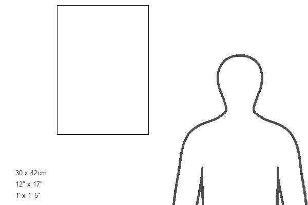
Estimated Image Size (if not cropped) is 29.7cm x 21cm (11.7" x 8.3")
Estimated Product Size is 29.7cm x 42cm (11.7" x 16.5")
These are individually made so all sizes are approximate
Artwork printed orientated as per the preview above, with landscape (horizontal) orientation to match the source image.
EDITORS COMMENTS
This print showcases Hartmannella vermiformis protozoa cysts, providing a glimpse into the microscopic world of single-celled organisms. Taken using a transmission electron microscope (TEM), the image reveals round cysts containing these fascinating creatures. H. vermiformis is commonly found in soil and freshwater environments, where it thrives by feeding on bacteria. The intricate details captured in this photograph highlight the biological complexity of these unicellular parasites. The sectioned cysts exhibit an abnormality caused by infection, offering valuable insights into their life cycle and potential impact on other organisms. With its histological significance, this image holds great importance for various fields such as zoology, biology, medicine, and microbiology. Researchers can utilize it to study the structure and behavior of H. vermiformis protozoa in order to better understand their role as infectious agents. Beyond its scientific value, this print also appeals to nature enthusiasts who appreciate wildlife photography at all scales – from majestic animals to microorganisms that play crucial roles in ecosystems. AMI IMAGES/SCIENCE PHOTO LIBRARY has expertly captured this mesmerizing snapshot of Hartmannella vermiformis protozoa cysts through meticulous microscopy techniques. As we delve deeper into the intricacies of our natural world, images like these continue to inspire awe and expand our knowledge about the diverse forms of life that surround us every day.
MADE IN THE UK
Safe Shipping with 30 Day Money Back Guarantee
FREE PERSONALISATION*
We are proud to offer a range of customisation features including Personalised Captions, Color Filters and Picture Zoom Tools
SECURE PAYMENTS
We happily accept a wide range of payment options so you can pay for the things you need in the way that is most convenient for you
* Options may vary by product and licensing agreement. Zoomed Pictures can be adjusted in the Basket.