Fraud Blocker Skip to main content

Protozoan Collection

"Exploring the Microscopic World: Unveiling the Diversity Life" In this captivating image series

Background imageProtozoan Collection: Acrosphaera radiolarian, SEM

Acrosphaera radiolarian, SEM
Acrosphaera radiolarian. Coloured scanning electron micrograph (SEM) of the shell of a Acrosphaera sp. radiolarian. Radiolaria are single-celled protozoans that are found in marine plankton

Background imageProtozoan Collection: Calcareous phytoplankton fossil, SEM Z100 / 0213

Calcareous phytoplankton fossil, SEM Z100 / 0213
Calcareous phytoplankton fossil. Coloured scanning electron micrograph (SEM) of a fossil of the skeleton (coccosphere) of a coccolithotrope, a small marine algal organism

Background imageProtozoan Collection: Trypanosome protozoan, artwork

Trypanosome protozoan, artwork
Trypanosome protozoan. Artwork of a section through a Trypanosoma congolense protozoan. This parasite is a cause of the disease nagana in cattle and other livestock in Africa

Background imageProtozoan Collection: Plasmodium sp. malarial parasite

Plasmodium sp. malarial parasite
Scanning electron microscope image of a malarial protozoal parasite. The parasite requires the anopheles mosquito to complete its life cycle

Background imageProtozoan Collection: Protozoa, a single-celled organism that feeds by scavenging for particles

Protozoa, a single-celled organism that feeds by scavenging for particles and other microorganisms, such as bacteria, or by absorbing nutrients from their environment

Background imageProtozoan Collection: Sleeping sickness parasite

Sleeping sickness parasite
Sleeping sickness. Artwork of a trypanosome (Trypanosoma brucei) moving past human red blood cells in the blood. This protozoan is the cause of sleeping sickness (African trypanosomiasis)

Background imageProtozoan Collection: Diatoms

Diatoms
Selected slide of a group of fossil diatoms collected from Bori, Hungary in September 1895 and viewed under the light microscipe using differential interfereance contrast

Background imageProtozoan Collection: Dinoflagellate plankton, SEM

Dinoflagellate plankton, SEM
Dinoflagellate plankton. Coloured scanning electron micrograph (SEM) of the calcareous (calcium-based) test (shell) of a dinoflagellate Calciodinellum sp

Background imageProtozoan Collection: Foraminiferan test (shell) SEM

Foraminiferan test (shell) SEM
Foraminiferan test or shell (Elphidium crispum) lateral view, coloured scanning electron micrograph (SEM). Foraminifera are single-celled protozoa which construct

Background imageProtozoan Collection: Dinoflagellates, SEM

Dinoflagellates, SEM
Dinoflagellates. Coloured scanning electron micrograph (SEM) of Prorocentrum dinoflagellates (round) and Ceratium dinoflagellates (branched). Dinoflagellates are unicellular protozoans

Background imageProtozoan Collection: Oxytricha ciliate protozoan, SEM C019 / 0253

Oxytricha ciliate protozoan, SEM C019 / 0253
Oxytricha sp. ciliate protozoan, coloured scanning electron micrograph (SEM). Oxytricha is a tiny single-celled aquatic organism

Background imageProtozoan Collection: Foraminifer model

Foraminifer model
Model of typical nummulitic foraminfer after Zittel

Background imageProtozoan Collection: Mouse malaria parasite, SEM

Mouse malaria parasite, SEM
Mouse malaria parasite. Coloured scanning electron micrograph (SEM) of a Plasmodium berghei protozoan (yellow) and red blood cells. P. berghei is the parasite that causes malaria in mice

Background imageProtozoan Collection: Radiolaria (engraving)

Radiolaria (engraving)
5216884 Radiolaria (engraving) by German School, (19th century); Private Collection; (add.info.: Radiolaria. Illustration from Meyers Konversations-Lexicon (Bibliographisches Institut)

Background imageProtozoan Collection: Giardia lamblia protozoa, micrograph

Giardia lamblia protozoa, micrograph
Giardia lamblia protozoa, light micrograph. These single-celled parasitic protozoa infect the intestinal tract and are most common in tropical regions

Background imageProtozoan Collection: Amoeba proteus, amoebae

Amoeba proteus, amoebae
A glass model of amoebae, created by Leopold and Rudolf Blaschka in the late nineteenth century and held at the Natural History Museum, London

Background imageProtozoan Collection: LM of Vorticella ciliates on a green alga

LM of Vorticella ciliates on a green alga
Light micrograph of a group of Vorticella (small tethered balloons) adhering to the freshwater green alga Hydrodictyon (large green cells)

Background imageProtozoan Collection: Paramecium sp. protozoan, SEM

Paramecium sp. protozoan, SEM
Paramecium protozoan. Coloured scanning electron micrograph (SEM) of a Paramecium sp. protozoan. This single-celled organism is aquatic, living in freshwater habitats

Background imageProtozoan Collection: Colour SEM of Podocyrtis cothurnata, a radiolarian

Colour SEM of Podocyrtis cothurnata, a radiolarian
Radiolarian. Coloured scanning electron micrograph (SEM) of the test (skeleton) of a Podocyrtis cothurnata, a type of radiolarian protozoan

Background imageProtozoan Collection: Euglena gracilis, SEM

Euglena gracilis, SEM
Euglena gracilis protists. Coloured scanning electron micrograph (SEM) of Euglena gracilis protists. These organisms possess a unique combination of plant and animal characters. Like the algae, E

Background imageProtozoan Collection: Foraminifer

Foraminifer
Scanning electron microscope (SEM) image of a foraminifer - a single celled organism

Background imageProtozoan Collection: Light micrograph of a group of Euglena gracilis

Light micrograph of a group of Euglena gracilis
Euglena gracilis. Light micrograph of a group of the single celled protozoa, Euglena gracilis. Euglenoids are common in ponds and stagnant fresh- water

Background imageProtozoan Collection: LM of the marine dinoflagellate, Ceratium sp

LM of the marine dinoflagellate, Ceratium sp
Dinoflagellate. Light micrograph of the marine dinoflagellate Ceratium. Found in both freshwater and saltwater, dinoflagellates are single-celled protozoa that float in the water column as plankton

Background imageProtozoan Collection: Euglena gracilis

Euglena gracilis protists. Light micrograph of a group of Euglena gracilis protists. These organisms possess a unique combination of plant and animal characters. Like the algae, E

Background imageProtozoan Collection: Computer art of radiolarians (from Ernst Haeckel)

Computer art of radiolarians (from Ernst Haeckel)
Radiolarians. Computer-enhanced engraving of radiolarian species. These one-celled protozoan animals are found in marine plankton

Background imageProtozoan Collection: Paramecium bursaria protozoan, micrograph

Paramecium bursaria protozoan, micrograph
Paramecium bursaria protozoan, light micrograph. This ciliate protozoan inhabits freshwater, where it feeds mainly on bacteria

Background imageProtozoan Collection: Paramecium protozoa, light micrograph

Paramecium protozoa, light micrograph
Paramecium bursaria protozoa, light micrograph. These ciliate protozoa inhabit freshwater, where they feed mainly on bacteria

Background imageProtozoan Collection: Trichodina parasite, light micrograph

Trichodina parasite, light micrograph
Trichodina parasite. Differential interference contrast micrograph of Trichodina pediculus. This ciliate parasite lives on the skin, gills and mucous membranes of many aquatic animals

Background imageProtozoan Collection: Dorataspis diodon, radiolarian

Dorataspis diodon, radiolarian
A glass model of a radiolarian, created by Leopold and Rudolf Blaschka in the late nineteenth century and held at the Natural History Museum, London

Background imageProtozoan Collection: Aulacantha scolymantha, radiolarian

Aulacantha scolymantha, radiolarian
A glass model of a radiolarian, created by Leopold and Rudolf Blaschka in the late nineteenth century and held at the Natural History Museum, London

Background imageProtozoan Collection: Lycogala epidendrum, Wolfs Milk

Lycogala epidendrum, Wolfs Milk
Watercolour on paper, c.1838 by Anna Russell (nee Worsley) (1807-1876). Held in the Library and Archives Date: circa 1838

Background imageProtozoan Collection: Eucyrtidium cranoides, radiolarian

Eucyrtidium cranoides, radiolarian
A glass model of a radiolarian, created by Leopold and Rudolf Blaschka in the late nineteenth century and held at the Natural History Museum, London

Background imageProtozoan Collection: Actinophrys sol, heliozoan

Actinophrys sol, heliozoan
A glass model of a radiolarian, created by Leopold and Rudolf Blaschka in the late nineteenth century and held at the Natural History Museum, London

Background imageProtozoan Collection: Heliosphaera actinota, radiolarian

Heliosphaera actinota, radiolarian
A glass model of a radiolarian, created by Leopold and Rudolf Blaschka in the late nineteenth century and held at the Natural History Museum, London

Background imageProtozoan Collection: Aulosphaera elegantissima, radiolarian

Aulosphaera elegantissima, radiolarian
A glass model of a radiolarian, created by Leopold and Rudolf Blaschka in the late nineteenth century and held at the Natural History Museum, London

Background imageProtozoan Collection: Difflugia pyriformis, amoebae

Difflugia pyriformis, amoebae
A glass model of amoebae, created by Leopold and Rudolf Blaschka in the late nineteenth century and held at the Natural History Museum, London

Background imageProtozoan Collection: Fossils of algae, plants, insects and protozoa

Fossils of algae, plants, insects and protozoa.. Chromolithograph from Dr. Fr. Rolles Geology and Paleontology section in Gotthilf Heinrich von Schuberts Natural History, Schreiber, Munich, 1886

Background imageProtozoan Collection: Vorticella convallaria, protozoan

Vorticella convallaria, protozoan.. Handcolored copperplate engraving from George Shaw and Frederick Nodders The Naturalists Miscellany, London, 1797

Background imageProtozoan Collection: Unicellular eukaryotic organisms

Unicellular eukaryotic organisms
Microscopic Organisms Antique Print, Vintage Lithograph, Urtiere Protozoen, Protozoa Old Zoology Illustration Unicellular Microbiology

Background imageProtozoan Collection: Illustration of Didinium on its own and swallowing a Paramecium

Illustration of Didinium on its own and swallowing a Paramecium

Background imageProtozoan Collection: Illustration of reproduction cycle of slime mould (protist)

Illustration of reproduction cycle of slime mould (protist)

Background imageProtozoan Collection: Cross section biomedical illustration on grid of Worm, Fungi, Protozoa

Cross section biomedical illustration on grid of Worm, Fungi, Protozoa, and Bacteria infection and infestation

Background imageProtozoan Collection: Egypt, Eastern Saharan Libyan Desert, Qattara Depression, Protozoan nummulites

Egypt, Eastern Saharan Libyan Desert, Qattara Depression, Protozoan nummulites
Egypt. Eastern Saharan Libyan Desert. Qattara Depression. Protozoan nummulites

Background imageProtozoan Collection: Foraminiferan fossil, SEM

Foraminiferan fossil, SEM
Foraminiferan fossil. Coloured scanning electron micrograph (SEM) of the fossilised shell (test) of a foraminiferan. Foraminifera are single-celled marine protozoa that construct

Background imageProtozoan Collection: LM of the protozoan Paramecium sp

LM of the protozoan Paramecium sp
Light micrograph of the ciliate protozoan, Paramecium sp. They are one of the most highly- evolved of all unicellular animals, and are plentiful in fresh water ponds

Background imageProtozoan Collection: Coloured SEM of foraminiferan, Peneroplis sp

Coloured SEM of foraminiferan, Peneroplis sp
Foraminiferan shell. Coloured scanning electron micrograph (SEM) of the test (shell) of the Mediterranean foraminiferan, Peneroplis proteus

Background imageProtozoan Collection: SEM of Paramecium

SEM of Paramecium
Paramecium. Coloured scanning electron micrograph (SEM) of the single-celled protozoan, Paramecium caudatum. This ciliate animal has cilia on its cell membrane surface enabling it to swim with an

Background imageProtozoan Collection: Chilodonella ciliate protozoan, SEM

Chilodonella ciliate protozoan, SEM
Chilodonella ciliate protozoa, coloured scanning electron micrograph (SEM). These tiny single- celled organisms are found in fresh water


All Professionally Made to Order for Quick Shipping

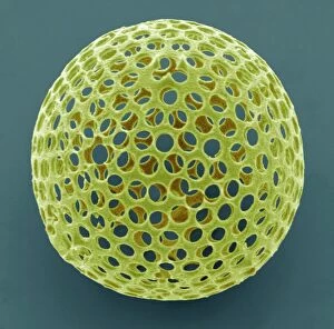
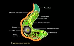
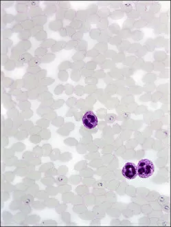
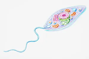
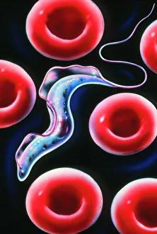
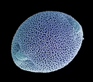
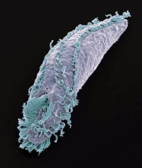
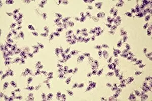
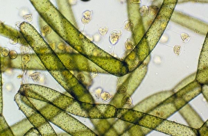
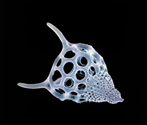
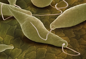
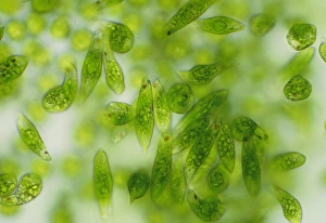
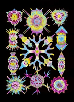
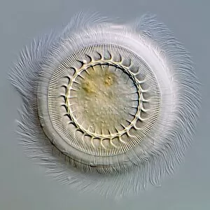
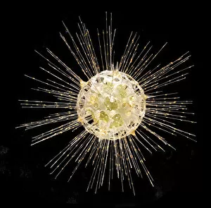
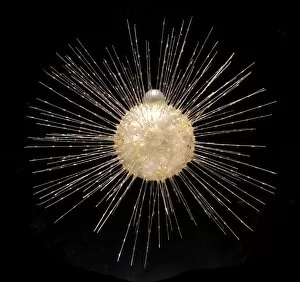
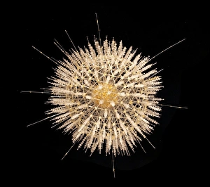
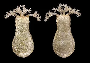
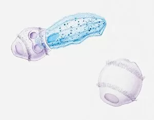
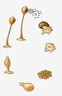
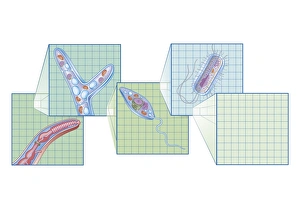
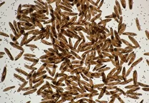
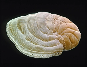
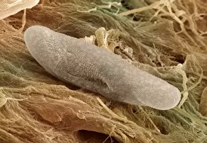
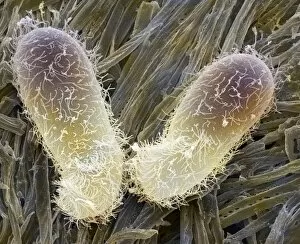
"Exploring the Microscopic World: Unveiling the Diversity Life" In this captivating image series, we delve into the fascinating realm of protozoa – a diverse group of single-celled organisms that inhabit our planet. From ancient calcareous phytoplankton fossils to intricate SEM images, prepare to be amazed by their incredible forms and functions. Starting with a glimpse into the past, we encounter an exquisite fossilized specimen of calcareous phytoplankton. Preserved in time, it offers us a window into Earth's history and evolution. Moving forward, our attention is captured by an artistic representation of Trypanosome protozoan – a notorious parasite responsible for causing sleeping sickness. Its mesmerizing artwork reminds us of both its beauty and danger lurking within nature. Shifting gears towards human health concerns, we come across Plasmodium sp. , the malarial parasite that has plagued humanity for centuries. This microscopic culprit serves as a stark reminder of the ongoing battle against malaria worldwide. Protozoa are known for their unique feeding strategies; some scavenge particles and microorganisms like bacteria while others absorb nutrients from their surroundings. Their adaptability is showcased through stunning SEM images featuring diatoms - intricately patterned unicellular algae - and Acrosphaera radiolarian - delicate marine organisms with intricate skeletal structures. The exploration continues as we encounter another menacing parasite responsible for mouse malaria. Through high-resolution SEM imagery, its complex morphology comes to life before our eyes. Diving deeper into this hidden world reveals Oxytricha ciliate protozoan – an organism characterized by its hair-like projections called cilia. The detailed SEM image showcases its extraordinary structure in vivid detail. Finally, we marvel at the intricate design of Foraminiferan tests (shells) captured using scanning electron microscopy (SEM). These tiny shells serve as protective homes for these remarkable creatures living in aquatic environments.