
Micro Organisms Collection
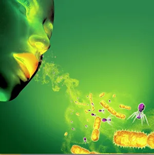
Microorganisms, the tiny wonders of life that exist all around us, are a fascinating subject to explore


All Professionally Made to Order for Quick Shipping
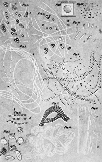
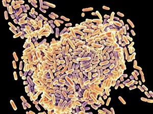
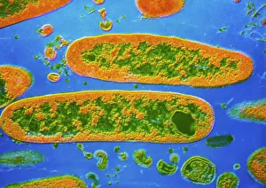
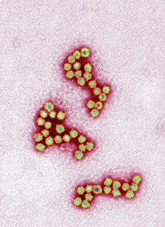
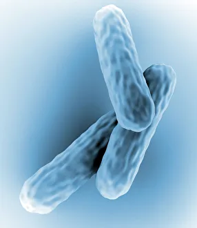
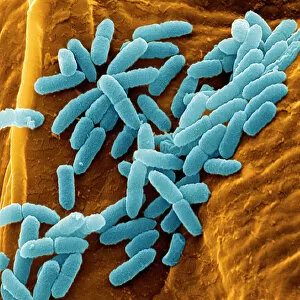
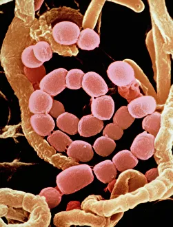
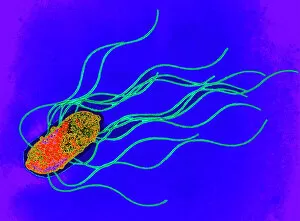
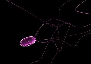
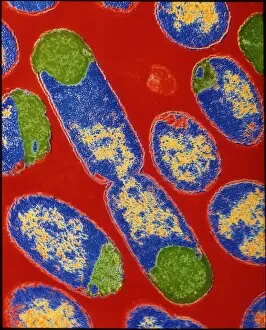
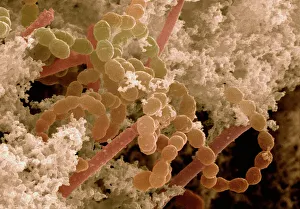
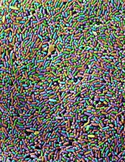
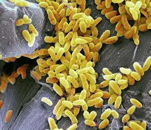
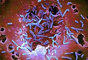
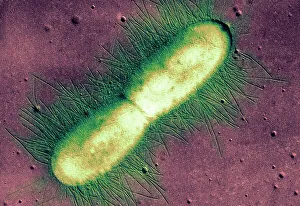
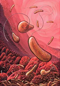
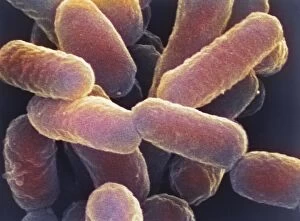
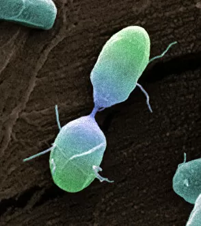
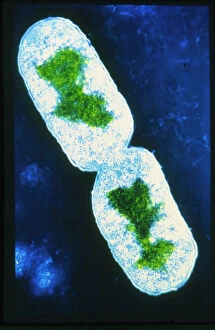
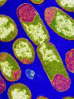
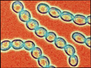
Microorganisms, the tiny wonders of life that exist all around us, are a fascinating subject to explore. Under the lens of a scanning electron microscope (SEM), we can witness their intricate structures and unravel their hidden secrets. Take E. Coli bacteria for example; when magnified through an SEM, they reveal their rod-shaped bodies with flagella protruding from one end. These microscopic creatures play crucial roles in our digestive system but can also cause infections if not properly handled. Similarly, Salmonella bacteria appear as elongated cells under SEM, reminding us of the importance of proper food handling and hygiene practices to prevent contamination. The colored transmission electron microscopy (TEM) image of Yersinia pestis bacteria showcases its unique features that were responsible for devastating outbreaks like the infamous Black Death. Switching gears to fungi, Candida fungus is captured beautifully in an SEM image displaying its filamentous structure. This opportunistic pathogen can cause infections in immunocompromised individuals and highlights the need for effective antifungal treatments. Delving into history, we encounter anthrax cultures depicted in a historical diagram. This bacterium has been weaponized throughout time due to its ability to form spores resistant to harsh conditions – a chilling reminder of humanity's dark side. Norovirus particles come into focus through TEM imagery; these small viral entities are notorious for causing gastroenteritis outbreaks worldwide and serve as a constant reminder about practicing good personal hygiene habits. Tuberculosis bacteria capture attention with their distinctive shape under SEM: slender rods often forming chains resembling delicate spirals. This ancient disease continues to pose significant health challenges globally despite medical advancements made over centuries. Streptomyces bacteria showcase their beauty by forming spiral spore chains visible even without high-powered microscopes. These remarkable organisms produce antibiotics vital for human health while maintaining ecological balance within soil ecosystems. Flagellate bacteria remind us that movement is not limited solely to larger organisms; these tiny creatures possess whip-like appendages that propel them through their microscopic habitats.