Fraud Blocker Skip to main content

Amoeboid Collection

"Exploring the Intricate World Creatures

Background imageAmoeboid Collection: Acrosphaera radiolarian, SEM

Acrosphaera radiolarian, SEM
Acrosphaera radiolarian. Coloured scanning electron micrograph (SEM) of the shell of a Acrosphaera sp. radiolarian. Radiolaria are single-celled protozoans that are found in marine plankton

Background imageAmoeboid Collection: Foraminifera, SEM

Foraminifera, SEM
Foraminifera. Coloured scanning electron micrograph (SEM) of a selection of foraminiferans, including Oolina hexagona (bottom right) and a number of Lagena sp

Background imageAmoeboid Collection: Plate 1 Circogonia Phaeodaria from Kunstformen der Natur (Art Forms in Nature)

Plate 1 Circogonia Phaeodaria from Kunstformen der Natur (Art Forms in Nature) illustrated by Ernst Haeckel (1834-1919)
5615747 Plate 1 Circogonia Phaeodaria from Kunstformen der Natur (Art Forms in Nature) illustrated by Ernst Haeckel (1834-1919) by Haeckel

Background imageAmoeboid Collection: Conceptual image of Radiolarians with a skeletal frame

Conceptual image of Radiolarians with a skeletal frame. Radiolarians are tiny protozoans that live in the ocean

Background imageAmoeboid Collection: Cancer cell with red blood cell flow

Cancer cell with red blood cell flow

Background imageAmoeboid Collection: Conceptual image of cancer virus

Conceptual image of cancer virus

Background imageAmoeboid Collection: 3D rendering of macrophage phagocytosis

3D rendering of macrophage phagocytosis

Background imageAmoeboid Collection: Microscopic view of phagocytic macrophages

Microscopic view of phagocytic macrophages, which are involved in the immune response within the body

Background imageAmoeboid Collection: Microscopic view of cancer virus

Microscopic view of cancer virus

Background imageAmoeboid Collection: Isolated cancer cell Macrophage

Isolated cancer cell Macrophage. Macrophages contribute to tumor growth and progression. Attracted to oxygen-starved (hypoxic) and necrotic tumor cells they promote chronic inflammation

Background imageAmoeboid Collection: Microscopic view of cancer cells

Microscopic view of cancer cells. Cancer occurs when a cells gene mutations make the cell unable to correct DNA damage

Background imageAmoeboid Collection: Hartmannella vermiformis protozoa cysts C016 / 9402

Hartmannella vermiformis protozoa cysts C016 / 9402
Hartmannella vermiformis protozoa cysts. Transmission electron micrograph (TEM) of a section through cysts (round) containing Hartmannella vermiformis protozoa. H

Background imageAmoeboid Collection: Amoeboid protozoa, SEM

Amoeboid protozoa, SEM
Amoeboid protozoa. Differential interference contrast (DIC) micrograph of four Thecamoeba sp. amoebozoa. Amoebozoa are a major group of amoeboid protozoa, motile single-celled organisms

Background imageAmoeboid Collection: Radiolarian, SEM C014 / 4862

Radiolarian, SEM C014 / 4862
Radiolarian. Coloured scanning electron micrograph (SEM) of the shell or test of a radiolarian. Radiolaria are single-celled protozoans that are found in marine plankton

Background imageAmoeboid Collection: Radiolarian, SEM C014 / 4863

Radiolarian, SEM C014 / 4863
Radiolarian. Coloured scanning electron micrograph (SEM) of the shell or test of a radiolarian. Radiolaria are single-celled protozoans that are found in marine plankton

Background imageAmoeboid Collection: Radiolaria, light micrograph C014 / 4672

Radiolaria, light micrograph C014 / 4672
Radiolaria. Collection of light micrographs showing various different species of radiolaria. Radiolaria (or radiolarians) are amoeboid protozoa that produce intricate mineral skeletons

Background imageAmoeboid Collection: Malaria parasite in red blood cell C016 / 2444

Malaria parasite in red blood cell C016 / 2444
Malaria parasite in red blood cell. Light micrograph of a red blood cell (erythrocyte) infected with a Plasmodim vivax protozoan. This parasite causes recurring malaria in humans

Background imageAmoeboid Collection: Malaria parasite in red blood cell C016 / 2442

Malaria parasite in red blood cell C016 / 2442
Malaria parasite in red blood cell. Light micrograph of a red blood cell (erythrocyte) infected with a Plasmodim vivax protozoan. This parasite causes recurring malaria in humans

Background imageAmoeboid Collection: Foraminiferan, SEM

Foraminiferan, SEM
Foraminiferan. Coloured scanning electron micrograph (SEM) of the foraminiferan Oolina squamosa. Foraminiferans are marine single-celled protists that construct and inhabit shells (tests)

Background imageAmoeboid Collection: Orbulina foraminiferan, SEM

Orbulina foraminiferan, SEM
Orbulina. Coloured scanning electron micrograph (SEM) of the shell of the foraminiferan Orbulina sp. Foraminiferans are marine single-celled protists that construct and inhabit shells (tests)

Background imageAmoeboid Collection: Naegleria fowleri protozoa, TEM

Naegleria fowleri protozoa, TEM
Naegleria fowleri protozoan. Coloured transmission electron micrograph (TEM) of a section through a Naegleria fowleri protozoan

Background imageAmoeboid Collection: Lychnocanium radiolarian, SEM

Lychnocanium radiolarian, SEM
Lychnocanium radiolarian. Coloured scanning electron micrograph (SEM) of the shell of a Lychnocanium sp. radiolarian. Radiolaria are single-celled protozoans that are found in marine plankton

Background imageAmoeboid Collection: Naegleria fowleri protozoan, TEM

Naegleria fowleri protozoan, TEM
Naegleria fowleri protozoan. Coloured transmission electron micrograph (TEM) of a section through a Naegleria fowleri protozoan

Background imageAmoeboid Collection: Amphisphaerina radiolarian, SEM

Amphisphaerina radiolarian, SEM
Amphisphaerina radiolarian. Coloured scanning electron micrograph (SEM) of the shell of a Amphisphaerina sp. radiolarian. Radiolaria are single-celled protozoans that are found in marine plankton

Background imageAmoeboid Collection: Acrosphaera radiolarian, SEM

Acrosphaera radiolarian, SEM
Acrosphaera radiolarian. Coloured scanning electron micrograph (SEM) of the shell of a Acrosphaera sp. radiolarian. Radiolaria are single-celled protozoans that are found in marine plankton

Background imageAmoeboid Collection: Foraminiferans, light micrograp

Foraminiferans, light micrograp
Foraminiferans, polarised light micrograph. Foraminiferans are marine single-celled protists that construct and inhabit shells (tests)

Background imageAmoeboid Collection: Radiolarian, SEM

Radiolarian, SEM
Radiolarian. Coloured scanning electron micrograph (SEM) of the shell of a radiolarian. Radiolaria are single-celled protozoans that are found in marine plankton

Background imageAmoeboid Collection: Radiolarian skeleton, artwork

Radiolarian skeleton, artwork
Radiolarian skeleton. Computer artwork of the skeleton (test) of a radiolarian. Radiolaria are single-celled protozoans that are found in marine plankton

Background imageAmoeboid Collection: Slime mould

Slime mould. Plasmodial stage of the myxomycete Physarum sp. on a dead treestump


All Professionally Made to Order for Quick Shipping

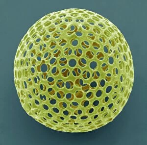
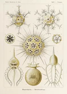
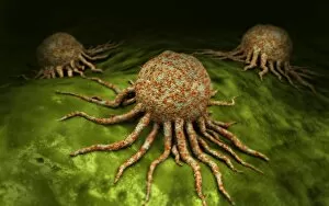
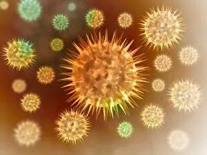
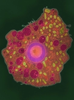
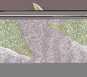
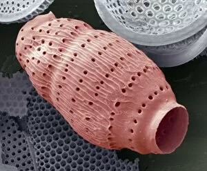
"Exploring the Intricate World Creatures: Acrosphaera radiolarian and Circogonia Phaeodaria" In Plate 1 of Ernst Haeckel's renowned Kunstformen der Natur (Art Forms in Nature), we are introduced to the mesmerizing beauty of Acrosphaera radiolarian. This intricate SEM image captures the delicate structure and unique characteristics of this amoeboid organism, showcasing its stunning complexity. Moving on to Picture No. 11675479, our fascination deepens as we encounter Circogonia Phaeodaria, another captivating example from Haeckel's collection. Its ethereal form seems almost otherworldly, leaving us in awe of nature's boundless creativity. As we delve further into the microscopic realm, a conceptual image reveals Radiolarians with their skeletal frames. These tiny creatures construct intricate structures that serve both as protection and support for their fragile bodies – a testament to their remarkable adaptability. Shifting gears slightly, we explore cancer cells through vivid imagery. In one snapshot, red blood cells flow alongside cancer cells, reminding us of the relentless battle waged within our own bodies against this formidable disease. Conceptual images depicting cancer viruses captivate our attention once again – these haunting visuals symbolize the invisible enemy that threatens human health worldwide. They serve as a stark reminder of the ongoing quest for effective treatments and cures in modern medicine. Microscopic views offer glimpses into phagocytic macrophages at work – tirelessly engulfing foreign particles and pathogens to protect our immune system from harm. Their tireless efforts underscore the importance of these crucial defenders within our bodies' defense mechanisms. Concluding our journey is a striking 3D rendering showcasing macrophage phagocytosis in action – an impressive display highlighting how these specialized cells efficiently eliminate threats by engulfing them entirely.