Glass Place Mat : Fractured atlas vertebra, 3D CT scan


![]()
Home Decor from Science Photo Library
Fractured atlas vertebra, 3D CT scan
Fractured atlas vertebra, coloured 3D CT (computed tomography) scan. Posterior view of a fractured atlas vertebra (break at upper centre) of a 34 year old man. Part of the skull can be seen at top. The atlas vertebra (or C1) is the topmost vertebra (spinal bone) that, along with the C2 vertebra, forms the joint connecting the skull and the spine. For a picture of a second fracture in the atlas vertebra of the same patient see M330/1706
Science Photo Library features Science and Medical images including photos and illustrations
Media ID 6424161
© DU CANE MEDICAL IMAGING LTD/SCIENCE PHOTO LIBRARY
Atlas Break Broken Cervical Computed Tomography Ct Scan Fracture Injured Injury Joint Neck Osteology Posterior Scanner Skeletal Spinal Column Thirties Three Dimensional Vertebra Vertebral Broken Neck Condition Disorder False Coloured Health Care Topmost Vertebrae
Glass Place Mat (Set of 4)
Set of 4 Glass Place Mats. Stylish and elegant polished safety glass, toughened and heat resistant (275x225mm, 7mm thick). Matching Coasters also available.
Set of 4 Glass Place Mats. Elegant polished safety glass and heat resistant. Matching Coasters may also be available
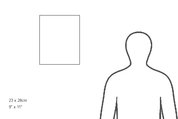
Estimated Image Size (if not cropped) is 22.5cm x 25.4cm (8.9" x 10")
Estimated Product Size is 22.5cm x 27.5cm (8.9" x 10.8")
These are individually made so all sizes are approximate
EDITORS COMMENTS
This print showcases a fractured atlas vertebra, captured in a vivid and detailed 3D CT scan. The image provides a posterior view of the injury, revealing a break at the upper center of the vertebra. At the top of the frame, we catch a glimpse of part of the skull, emphasizing the intricate connection between our skeletal structure and spinal column. The atlas vertebra, also known as C1, holds significant importance as it forms the joint that links our skull to our spine along with the C2 vertebra. This particular fracture belongs to a 34-year-old man whose condition is depicted here. It offers an intriguing insight into medical science and highlights both human anatomy and pathology. With its false-colored presentation, this photograph allows us to delve into osteology and gain valuable knowledge about fractures within our skeletal system. The three-dimensional aspect adds depth to our understanding by providing an immersive visual experience. As we explore this image further, we are reminded of how delicate yet resilient our bodies can be. It serves as a reminder of both fragility and strength while shedding light on medical advancements in health care technology such as CT scans or computed tomography.
MADE IN THE UK
Safe Shipping with 30 Day Money Back Guarantee
FREE PERSONALISATION*
We are proud to offer a range of customisation features including Personalised Captions, Color Filters and Picture Zoom Tools
SECURE PAYMENTS
We happily accept a wide range of payment options so you can pay for the things you need in the way that is most convenient for you
* Options may vary by product and licensing agreement. Zoomed Pictures can be adjusted in the Basket.