
Computed Tomography Collection
Computed tomography (CT) is a revolutionary medical imaging technique that has transformed the field of diagnostics


All Professionally Made to Order for Quick Shipping
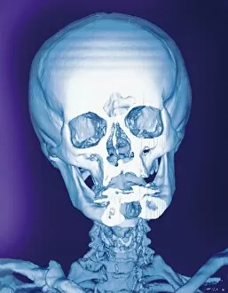
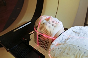
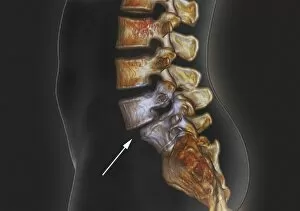
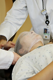
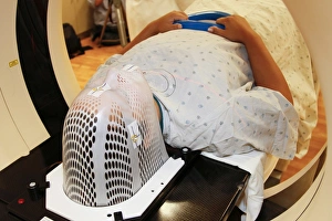
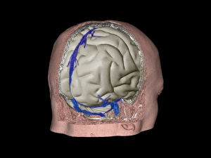
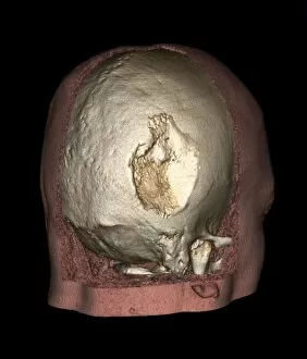
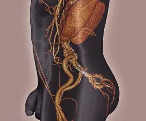
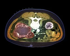
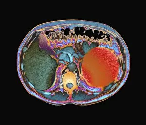
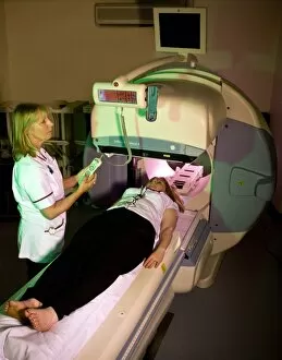
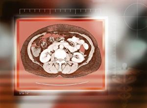
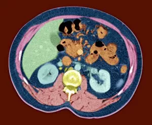
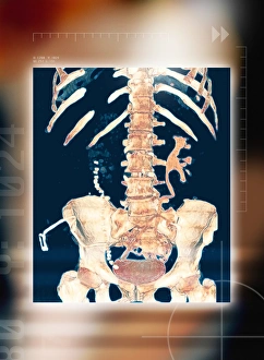
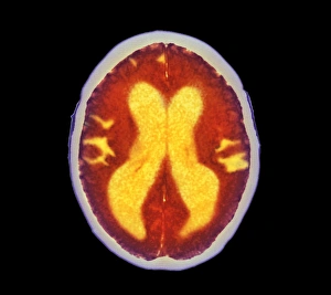
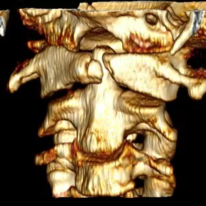
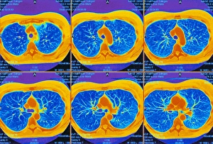
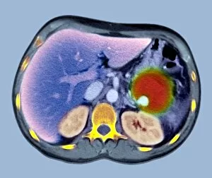
Computed tomography (CT) is a revolutionary medical imaging technique that has transformed the field of diagnostics. With its ability to provide detailed cross-sectional images, CT scans have become an indispensable tool for healthcare professionals. Starting with a normal skull, CT allows us to visualize intricate structures within our cranium that cannot be seen on a regular X-ray. From teeth anatomy illustrations to body imaging, this technology provides invaluable insights into various conditions and abnormalities. A side view of the neck in colored X-ray reveals the complexity of our cervical vertebrae. Just like a chrysanthemum's petals unfold beautifully, CT scan images unravel hidden details within our bodies. Lasers are used to precisely position patients wearing short face masks during computed tomography procedures. This ensures accurate image acquisition and minimizes any potential movement artifacts. Incredible 3D scans showcase the power of CT in diagnosing complex conditions such as brain aneurysms, spondylolisthesis, and aortic dissection. These high-resolution images enable doctors to plan interventions with precision and improve patient outcomes. Not limited to specific areas, CT can also produce comprehensive 3D scans of normal abdomens. This non-invasive technique aids in detecting diseases or anomalies affecting internal organs without resorting to surgery or invasive procedures. Radiation therapists play a crucial role by fitting short face masks on patients during radiation therapy sessions using guidance from CT scans. This personalized approach ensures targeted treatment while minimizing damage to healthy tissues surrounding the affected area. As patients are positioned inside the computed tomography scanner, they embark on a journey through cutting-edge technology that unveils their innermost secrets layer by layer and has revolutionized medical imaging by providing detailed visualizations beyond what traditional methods could achieve. Its applications range from dental anatomy illustrations and body imaging to diagnosing life-threatening conditions like brain aneurysms and spondylolisthesis – all while ensuring patient safety and comfort.