Home > Science > Xray
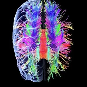
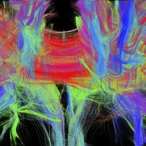
Brain aneurysm, 3D scan

![]()
Wall Art and Photo Gifts from Science Photo Library
Brain aneurysm, 3D scan
Brain aneurysm. 3D computed tomography (CT) angiogram coupled with a magnetic resonance imaging (MRI) scan of the brain of a 38-year-old, showing a large aneurysm (bright, lower centre) of the right internal carotid artery. An aneurysm is the ballooning of an artery due to weakening of the artery wall
Science Photo Library features Science and Medical images including photos and illustrations
Media ID 9247097
© ZEPHYR/SCIENCE PHOTO LIBRARY
38 Year Old 38 Years Old Aneurism Aneurysm Angiogram Angiography Arterial Arteries Bleed Bleeding Cerebral Cerebrovascular Colored Computed Tomography Cranial Diagnosis Diagnostic Imaging Haemorrhage Hemorrhage Magnetic Resonance Imaging Meninges Profile Radiography Radiological Radiology Thirties Vascular System Vessels Xray Abnormal Blood Vessel Brain Circulatory System Condition Disorder Unhealthy
FEATURES IN THESE COLLECTIONS
EDITORS COMMENTS
This print from Science Photo Library showcases a 3D scan of a brain aneurysm, providing valuable insights into this potentially life-threatening condition. The image features a vibrant and detailed display of the brain's circulatory system, with the focus on a large aneurysm located in the lower center of the right internal carotid artery. The black background enhances the visual impact, allowing us to fully appreciate the intricate network of arteries and blood vessels that make up our cerebral anatomy. This side view perspective offers a unique glimpse into the inner workings of our complex neurological structure. At just 38 years old, this individual's diagnosis highlights how aneurysms can affect individuals across all age groups. The bright coloring emphasizes the abnormality within their arterial system, caused by weakening in the artery wall leading to ballooning or swelling. With bleeding being one potential consequence of such conditions, it serves as a stark reminder of why early detection and medical intervention are crucial. This powerful image underscores both the fragility and resilience of our bodies while emphasizing advancements in diagnostic imaging techniques like magnetic resonance imaging (MRI) and computed tomography (CT). Science Photo Library continues to provide invaluable contributions to healthcare through their extensive collection capturing various medical conditions with scientific precision.
MADE IN THE UK
Safe Shipping with 30 Day Money Back Guarantee
FREE PERSONALISATION*
We are proud to offer a range of customisation features including Personalised Captions, Color Filters and Picture Zoom Tools
SECURE PAYMENTS
We happily accept a wide range of payment options so you can pay for the things you need in the way that is most convenient for you
* Options may vary by product and licensing agreement. Zoomed Pictures can be adjusted in the Basket.