
Vertebral Collection
"Exploring the Intricacies of the Vertebral Column: From Slipped Discs to Ancient Anatomy" The vertebral column, commonly known as the backbone


All Professionally Made to Order for Quick Shipping
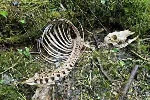
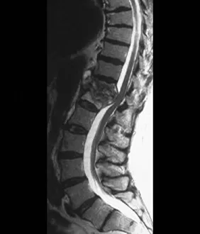
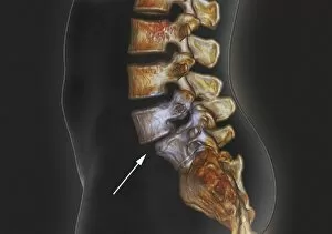
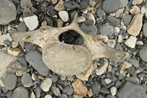
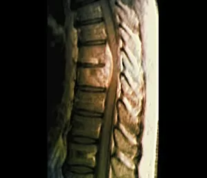
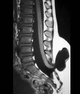
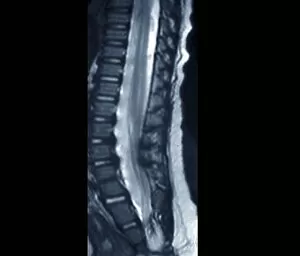
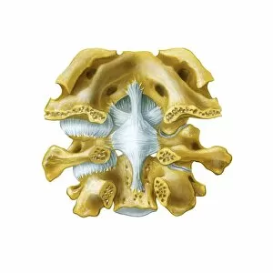
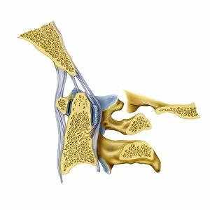
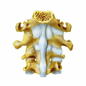
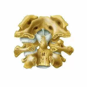
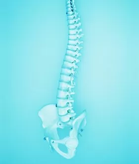
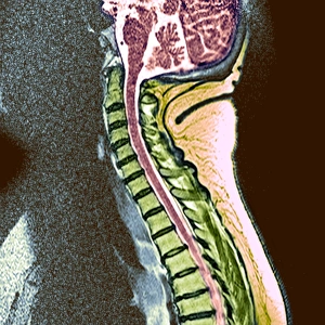
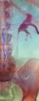
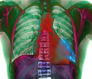
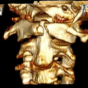
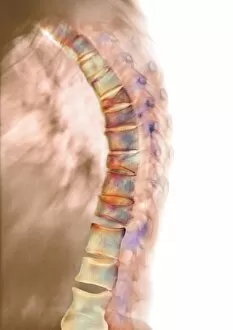
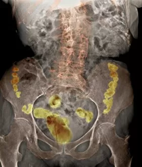
"Exploring the Intricacies of the Vertebral Column: From Slipped Discs to Ancient Anatomy" The vertebral column, commonly known as the backbone, is a fascinating structure that supports our bodies and protects our spinal cord. It plays a crucial role in maintaining our posture and facilitating movement. One common issue associated with the vertebral column is a slipped disc, which occurs when one of the intervertebral discs protrudes out of place. This condition can cause immense pain and discomfort but can be diagnosed through an MRI scan. Speaking of scans, modern medical technology allows us to delve deep into the intricacies of this vital part of our anatomy. MRI scans provide detailed images showcasing both normal torsos and abnormalities such as tuberculosis or spondylolisthesis affecting the spine. Art has also played a significant role in depicting head and neck anatomy throughout history. Stunning artwork showcases these intricate structures, capturing their beauty while providing educational insights. Comparisons between species are another intriguing aspect related to vertebrae. A lithograph comparing a man's skeletal structure with that of a gorilla highlights both similarities and differences between humans and primates. Furthermore, ancient remains like red deer skeletons or Australopithecus africanus pelvis (STS-14 C015 / 6919) offer valuable glimpses into how different species evolved over time. Eadweard Muybridge's iconic photograph "The Walk" captures human locomotion by freezing each step taken during walking—a testament to how every movement relies on proper functioning vertebrae. Lastly, let's not forget about the brain and spinal cord—the ultimate beneficiaries of well-maintained vertebrae. Artwork depicting these essential components reminds us just how interconnected they are within our bodies' complex systems. Exploring various aspects surrounding vertebral health provides us with invaluable knowledge about its importance for overall well-being.