Glass Frame > Science > SEM
Glass Frame : Collecting duct from a kidney, SEM


![]()
Mounted Prints from Science Photo Library
Collecting duct from a kidney, SEM
Collecting duct from a kidney. Coloured scanning electron micrograph (SEM) of a section through a collecting duct from a kidney. This section across the duct (a thin tube) shows a thick, folded wall surrounding a narrow central space (lumen). This collecting duct receives urine from the Loop of Henle (which is part of a kidney structure called a tubule). The urine is the product of the blood filtration that occurred elsewhere in the kidney. The Loop of Henle reabsorbs water and salts from the urine, increasing the concentration of waste products such as urea. Magnification: x12, 500 when printed 10cm wide
Science Photo Library features Science and Medical images including photos and illustrations
Media ID 6422746
© STEVE GSCHMEISSNER/SCIENCE PHOTO LIBRARY
Cross Section Duct Excretory System Histological Histology Human Biology Kidney Lumen Nephrology Renal Thick Transverse Tube Tubule Urine Urological Urology Wall False Coloured Section Sectioned
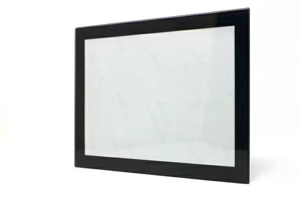
8"x6" Glass Mount
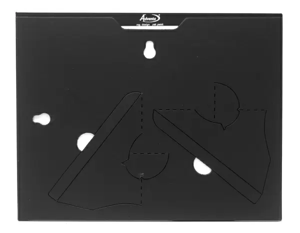
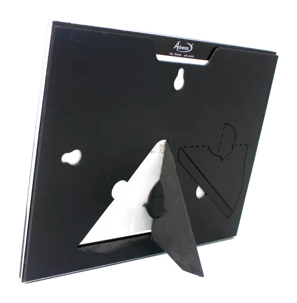
Wall mounted or free-standing, these black edged glass frames feature a smooth chamfered edge and a stylish black border (on back face of the glass). Manufactured from 4mm thick glass, Glass Mounts are a durable, professional way of displaying and protecting your prints. Your 8x6 print is slotted into the back of the frame so can easily be changed if needed.
Tempered Glass Mounts are ideal for wall display, plus the smaller sizes can also be used free-standing via an integral stand
Estimated Image Size (if not cropped) is 15.2cm x 20.3cm (6" x 8")
Estimated Product Size is 17.7cm x 22.8cm (7" x 9")
These are individually made so all sizes are approximate
Artwork printed orientated as per the preview above, with portrait (vertical) orientation to match the source image.
EDITORS COMMENTS
This print showcases a cross-section of a collecting duct from a kidney, revealing its intricate structure and function. The colored scanning electron micrograph (SEM) provides a detailed view of the thin tube surrounded by a thick, folded wall. Within this narrow central space known as the lumen, urine is received from the Loop of Henle. The Loop of Henle plays an essential role in renal physiology by reabsorbing water and salts from the urine, thereby increasing the concentration of waste products like urea. This process is crucial for maintaining proper fluid balance and eliminating toxins from our bodies. With a magnification level of x12,500 when printed at 10cm wide, this image offers an up-close look at the healthy anatomy of this vital component within our excretory system. Its false-colored presentation adds to its aesthetic appeal while still providing valuable scientific information. Whether you are studying human biology or exploring nephrology and urology fields, this histological sectioned image serves as an invaluable resource. It highlights the complexity and beauty found within our own bodies while shedding light on how they efficiently carry out essential functions like urine production and filtration. Captured by Science Photo Library using advanced scanning electron microscope technology, this photograph exemplifies their commitment to delivering high-quality images that bridge science with artistry.
MADE IN THE UK
Safe Shipping with 30 Day Money Back Guarantee
FREE PERSONALISATION*
We are proud to offer a range of customisation features including Personalised Captions, Color Filters and Picture Zoom Tools
SECURE PAYMENTS
We happily accept a wide range of payment options so you can pay for the things you need in the way that is most convenient for you
* Options may vary by product and licensing agreement. Zoomed Pictures can be adjusted in the Basket.