Fraud Blocker Skip to main content

Renal Collection

Renal, the intricate masterpiece of our cardiovascular system, has long been celebrated in historical artwork

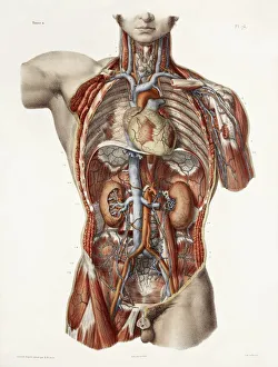
Background imageRenal Collection: Cardiovascular system, historical artwork

Cardiovascular system, historical artwork. 19th Century hand coloured lithographic print showing the blood vessels (red and blue) of the human neck and torso

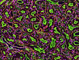
Background imageRenal Collection: Kidney tubules in section

Kidney tubules in section
Kidney tubules. Fluorescent light micrograph of a section through kidney tissue showing numerous tubules (black/green). The tubules are seen in cross-section

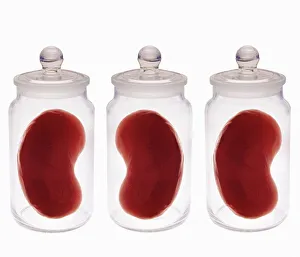
Background imageRenal Collection: Spare kidneys

Spare kidneys. Conceptual computer artwork of spare kidneys stored in jars. This may represent the growth of human tissue to produce material for transplant surgery to replace defective or old body

Background imageRenal Collection: Kidney glomeruli, SEM

Kidney glomeruli, SEM
Kidney glomeruli, coloured scanning electron micrograph (SEM). The cells that usually surround the glomeruli have been removed to reveal the coiled capillaries at the core of the glomeruli

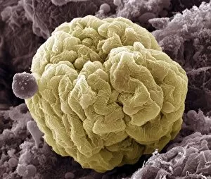
Background imageRenal Collection: Kidney glomerulus, SEM

Kidney glomerulus, SEM
Kidney glomerulus, coloured scanning electron micrograph (SEM). Podocyte cells, which make up the surface of the glomerulus, are yellow. Magnification: x650 when printed at 10 centimetres wide

Background imageRenal Collection: Kidney dialysis

Kidney dialysis
MODEL RELEASED. Kidney dialysis

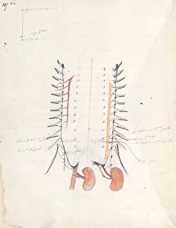
Background imageRenal Collection: The Kidneys, c. 1843 (pen & ink and coloured pencil on paper)

The Kidneys, c. 1843 (pen & ink and coloured pencil on paper)
CHT234657 The Kidneys, c.1843 (pen & ink and coloured pencil on paper) by Bernard, Claude (1813-78); Bibliotheque du College de France, Paris, France; eArchives Charmet; French, out of copyright

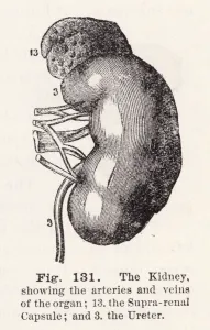
Background imageRenal Collection: The kidney, showing the arteries and veins of the organ (engraving)

The kidney, showing the arteries and veins of the organ (engraving)
2799336 The kidney, showing the arteries and veins of the organ (engraving) by English School, (19th century); Private Collection; (add.info.: The kidney, showing the arteries and veins of the organ)

Background imageRenal Collection: Female anatomy, historical model

Female anatomy, historical model
Female anatomy. 1920s anatomical model showing the internal organs of the female upper body with the intestines, stomach and liver removed

Background imageRenal Collection: Female abdominal anatomy, artwork

Female abdominal anatomy, artwork
Female abdominal anatomy. Computer artwork showing the kidneys and female reproductive system. Part of the lymphatic system (green) is also shown

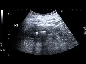
Background imageRenal Collection: Kidney stones, ultrasound scan C017 / 7381

Kidney stones, ultrasound scan C017 / 7381
Kidney stones. Ultrasound scan of a 83 year old female patient with two kidney stones (bright white) in the right kidney. Kidney stones, or calculi, are formed when mineral salts

Background imageRenal Collection: Polycystic kidney, ultrasound scan C017 / 7746

Polycystic kidney, ultrasound scan C017 / 7746
Polycystic kidney. Ultrasound scan of the kidney of a 42 year old female showing numerous fluid-filled cysts (dark areas)

Background imageRenal Collection: Normal abdomen, 3D CT scan

Normal abdomen, 3D CT scan
Normal abdomen. Coloured 3D computed tomography (CT) scan of the healthy abdomen and chest of a 53 year old patient. At top centre is the heart

Background imageRenal Collection: Kidney anatomy, artwork

Kidney anatomy, artwork
Kidney anatomy. Cutaway artwork showing the internal structure of a kidney. The kidney, one of a pair of such organs, is part of the human excretory system

Background imageRenal Collection: Nephron structure, artwork

Nephron structure, artwork
Nephron structure. Artwork showing the structure of a nephron, the functional filtering unit of a kidney. The function of the kidney is to filter waste products such as urea from the blood

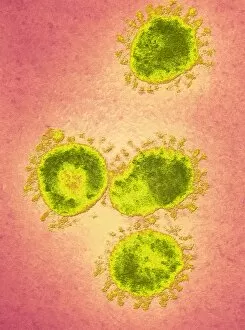
Background imageRenal Collection: Infectious bronchitis virus (IBV), TEM

Infectious bronchitis virus (IBV), TEM
Infectious bronchitis virus (IBV) particles, coloured transmission electron micrograph (TEM). This virus is a member of the coronavirus family

Background imageRenal Collection: Mitochondria, SEM

Mitochondria, SEM
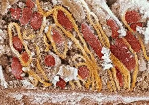
Mitochondria. Coloured scanning electron micrograph (SEM) of mitochondria (red) in a kidney cell. Mitochondria are a type of organelle found in the cytoplasm of eukaryotic cells

Background imageRenal Collection: Polycystic kidneys, ultrasound scan

Polycystic kidneys, ultrasound scan
Polycystic kidneys. Ultrasound scan of the kidney of an 82 year old male showing numerous fluid-filled cysts (dark areas)

Background imageRenal Collection: Healthy kidney, artwork

Healthy kidney, artwork
Healthy kidney, computer artwork

Background imageRenal Collection: Kidney anatomy, 1825 artwork

Kidney anatomy, 1825 artwork
Kidney anatomy. At top are the left and right kidneys (brown) with arteries (red), veins (blue), ureters (white) and adrenal glands (cream, on top of kidneys)

Background imageRenal Collection: Kidneys, anatomical artwork

Kidneys, anatomical artwork
Kidneys. Anatomical computer artwork of a mans body, showing the position of the kidneys (red, centre left and right) and adrenal glands (beige)

Background imageRenal Collection: Atherosclerosis, 3D CT scan

Atherosclerosis, 3D CT scan
Atherosclerosis. 3D computed tomography (CT) angiogram of the main blood vessels of the lower body a 58-year-old man, showing numerous atheromatous plaques (light, centre) on the vessel walls

Background imageRenal Collection: Kidney anatomy, artwork C016 / 8945

Kidney anatomy, artwork C016 / 8945
Kidney anatomy, artwork. The kidneys filter waste from the blood and excrete it as urine. The kidney consists of an outer cortex and an inner medulla

Background imageRenal Collection: Incontinence surgery, artwork C016 / 6843

Incontinence surgery, artwork C016 / 6843
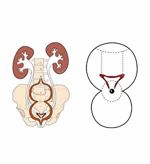
Incontinence surgery. Artwork of the sacral region of a womans pelvis showing a schematic diagram for bladder and sacral surgery to treat incontinence

Background imageRenal Collection: Inflamed kidney

Inflamed kidney
Inflamed and swollen kidney, gross specimen. This is a case of chronic nephritis, an inflammation of the kidney. Nephritis is often caused by a bacterial infection

Background imageRenal Collection: Glomerulus, TEM

Glomerulus, TEM
Glomerulus. Transmission electron micrograph (TEM) of a section through a nephron in a human kidney, showing a cluster of capillaries within a renal corpuscle, also known as a glomerulus

Background imageRenal Collection: Organ transplants, conceptual artwork

Organ transplants, conceptual artwork
Organ transplants. Conceptual artwork of a human body as a jigsaw puzzle, with two removed puzzle pieces representing organ transplants

Background imageRenal Collection: Kidney mitochondria, TEM

Kidney mitochondria, TEM
Kidney mitochondria. Transmission electron micrograph (TEM) of a section through a kidney tubule, showing numerous mitochondria (blue)

Background imageRenal Collection: Kidney tumour, CT scan

Kidney tumour, CT scan
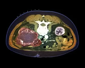
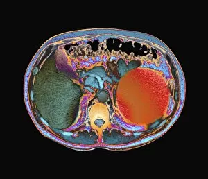
Kidney tumour. Coloured computed tomography (CT) scan of a section through the abdomen of a 62-year-old patient, showing necrosis (death) of tissue in the right kidney

Background imageRenal Collection: Polycystic kidney

Polycystic kidney. Gross specimen of a polycystic kidney, where most of the tissue of the kidneys has been replaced by cysts

Background imageRenal Collection: Polycystic kidneys, CT scans C018 / 0571

Polycystic kidneys, CT scans C018 / 0571
Polycystic kidneys. Coloured computed tomography (CT) scans of the abdomen of a 28 year old patient with polycystic kidneys (pink)

Background imageRenal Collection: Polycystic kidneys, CT scans C018 / 0572

Polycystic kidneys, CT scans C018 / 0572
Polycystic kidneys. Coloured computed tomography (CT) scans of the abdomen of a 28 year old patient with polycystic kidneys (dark blue)

Background imageRenal Collection: Polycystic kidneys, CT scan C018 / 0573

Polycystic kidneys, CT scan C018 / 0573
Polycystic kidneys. Coloured computed tomography (CT) scan of the abdomen of a 28 year old patient with polycystic kidneys (dark blue)

Background imageRenal Collection: Friedrich von Frerichs, German optician C018 / 7108

Friedrich von Frerichs, German optician C018 / 7108
Friedrich von Frerichs (1819-1885). 1889 engraving of the German ophthalmologist and pathologist Friedrich Theodor von Frerichs

Background imageRenal Collection: Nephron structure, artwork C016 / 9867

Nephron structure, artwork C016 / 9867
Nephron structure. Artwork showing the structure of a nephron, the functional filtering unit of a kidney. The function of the kidney is to filter waste products such as urea from the blood

Background imageRenal Collection: Nephron structure, artwork C016 / 9868

Nephron structure, artwork C016 / 9868
Nephron structure. Artwork showing the structure of a nephron, the functional filtering unit of a kidney. The function of the kidney is to filter waste products such as urea from the blood

Background imageRenal Collection: Healthy kidneys, X-ray C014 / 4920

Healthy kidneys, X-ray C014 / 4920
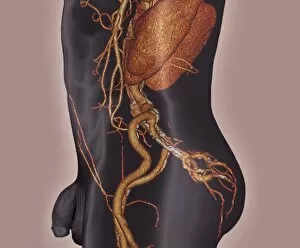
Healthy kidneys. Arteriograph (artery X-ray) of the abdomen showing two healthy kidneys (yellow, left and right). he kidneys filter waste products from the blood

Background imageRenal Collection: Healthy kidneys, X-ray C014 / 4921

Healthy kidneys, X-ray C014 / 4921
Healthy kidneys. Arteriograph (artery X-ray) of the abdomen showing two healthy kidneys (orange). he kidneys filter waste products from the blood

Background imageRenal Collection: Human kidneys, historical model

Human kidneys, historical model
Human kidneys. 1950s anatomical model of the human kidneys (red) with the ureters (tubes, upper left) attached, and a section through one kidney showing the internal structure

Background imageRenal Collection: Kidney anatomy, artwork C016 / 8663

Kidney anatomy, artwork C016 / 8663
Kidney anatomy, artwork. The kidneys filter waste from the blood and excrete it as urine. The kidney consists of an outer cortex and an inner medulla

Background imageRenal Collection: Kidney anatomy, artwork C016 / 8661

Kidney anatomy, artwork C016 / 8661
Kidney anatomy, artwork. The kidneys filter waste from the blood and excrete it as urine. The kidney consists of an outer cortex and an inner medulla

Background imageRenal Collection: Kidney anatomy, artwork C016 / 8662

Kidney anatomy, artwork C016 / 8662
Kidney anatomy, artwork. The kidneys filter waste from the blood and excrete it as urine. The kidney consists of an outer cortex and an inner medulla

Background imageRenal Collection: Kidney anatomy, artwork C016 / 8660

Kidney anatomy, artwork C016 / 8660
Kidney anatomy, artwork. The kidneys filter waste from the blood and excrete it as urine. The kidney consists of an outer cortex and an inner medulla

Background imageRenal Collection: Kidney tissue, confocal micrograph C014 / 4611

Kidney tissue, confocal micrograph C014 / 4611
Kidney tissue. Laser scanning confocal micrograph (LSCM) of a section through a human kidney, showing the cell nuclei (dark)

Background imageRenal Collection: Kidney tissue, fluorescence micrograph C016 / 8484

Kidney tissue, fluorescence micrograph C016 / 8484
Kidney tissue. Fluorescence deconvolution micrograph of a section through a kidney, showing glomeruli (green), cell nuclei (blue dots), and renal tubules (red, circular)

Background imageRenal Collection: Kidney blood vessels, confocal micrograph C014 / 4610

Kidney blood vessels, confocal micrograph C014 / 4610
Kidney blood vessels. Laser scanning confocal micrograph (LSCM) of a section through a human kidney, showing a blood vessel (vertical, centre)

Background imageRenal Collection: MDCK culture cells, SEM C016 / 8028

MDCK culture cells, SEM C016 / 8028
MDCK cells. Coloured scanning electron micrograph (SEM) of cultured MDCK (Madin-Darby canine kidney) epithelial cells on filter paper (fibres)

Background imageRenal Collection: Urinary system, artwork C015 / 6455

Urinary system, artwork C015 / 6455
Urinary system. Computer artwork showing the organs of the human urinary system. The kidneys (red, top left and right) filter waste products such as urea from the blood (red and blue) to form urine


All Professionally Made to Order for Quick Shipping

Renal, the intricate masterpiece of our cardiovascular system, has long been celebrated in historical artwork. From ancient paintings to modern sculptures, artists have attempted to capture the essence of this vital organ. When we delve deeper into its structure, we discover kidney tubules in section - a maze-like network responsible for filtering waste and maintaining fluid balance within our bodies. And if one is fortunate enough to possess spare kidneys, they serve as backup guardians of our health. The kidney glomerulus, captured magnificently through scanning electron microscopy (SEM), reveals its intricate web-like structure that allows it to efficiently filter blood and remove toxins. These mesmerizing images remind us of the beauty hidden within our own bodies. Abdominal arteries come alive in an X-ray image (P206 / 0309) showcasing their role in supplying oxygen-rich blood to the renal region. This snapshot highlights the interconnectedness between various components of our body's architecture. Kidney dialysis plays a crucial role for those whose renal function is compromised. It acts as a lifeline by artificially replicating the filtration process performed by healthy kidneys. This medical marvel ensures that individuals can continue living fulfilling lives despite their renal challenges. As we explore more SEM images capturing kidney glomeruli and blood vessels, we are reminded once again of nature's exquisite design. The delicate balance maintained within these structures reflects the harmony required for optimal functioning. Intriguingly juxtaposed with abdominal organs lies this artistic portrayal of kidneys - symbolic representations reminding us how essential they are for overall well-being. They silently work day and night behind-the-scenes performing countless tasks necessary for maintaining homeostasis within our bodies. Renal - not just another word but an embodiment of life-sustaining mechanisms intricately woven into every fiber of human existence. Let us appreciate this remarkable organ that tirelessly serves as a guardian protecting us from harm while quietly orchestrating numerous physiological processes essential for survival.