
Lumen Collection
"Lumen: Illuminating the Inner Workings of the Human Body" In the intricate realm of medical illustrations, lumen takes center stage as a captivating concept


All Professionally Made to Order for Quick Shipping
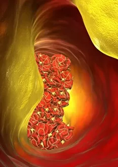
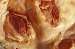
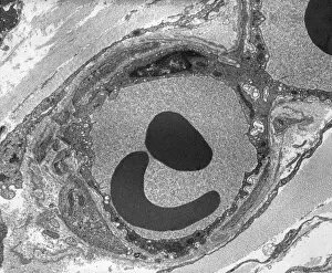
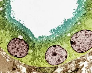
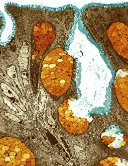
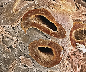
"Lumen: Illuminating the Inner Workings of the Human Body" In the intricate realm of medical illustrations, lumen takes center stage as a captivating concept. From an appendix plagued by appendicitis to intestinal villi viewed through a scanning electron microscope (SEM), these visual representations shed light on our innermost structures and their ailments. Delving deeper into this fascinating world, we encounter a fallopian tube captured under the lens of a light micrograph F006/9799. Its delicate intricacies remind us of the remarkable complexity involved in human reproduction. But it is not just organs that captivate our attention; even blood vessels have stories to tell. A computer artwork reveals a blocked artery, showcasing both its vulnerability and resilience. Picture No. 11675562 offers us insight into this critical aspect of cardiovascular health. As we explore further, Picture No. 11675561 beckons us with its enigmatic allure – what secrets lie within? The subsequent images, numbered from 11675560 to 11675555, invite curiosity and intrigue as they unveil various aspects of our internal landscape. Through these captivating visuals, lumen serves as more than just an anatomical term; it becomes a metaphor for enlightenment and understanding. It reminds us that beneath our skin lies an intricate network waiting to be explored – one that holds answers to countless mysteries surrounding human physiology. So let us embark on this journey together, guided by lumen's luminosity, unraveling the wonders hidden within each image until we gain profound insights into ourselves.