Glass Coaster : Knee injury, 3D CT scan C018 / 0639


![]()
Home Decor from Science Photo Library
Knee injury, 3D CT scan C018 / 0639
Knee injury. Coloured 3D computed tomography (CT) scan of a knee with a meniscal tear (indicated by arrows). The menisci are fibrocartilage crescents that act as shock absorbers between the femur (thigh bone, top) and tibia (shin bone, bottom). A tear in a meniscus can be caused by trauma, most often a sports injury, or by wear and tear. Treatment depends on the severity of the tear. Minor tears may respond to physical therapy and anti-inflammatory drugs, but more serious injuries require surgical treatment
Science Photo Library features Science and Medical images including photos and illustrations
Media ID 9237821
© ZEPHYR/SCIENCE PHOTO LIBRARY
Cartilage Ct Scan Damage Damaged Diagnosis Diagnostic Femur Fibrocartilage Injured Injury Joint Knee Meniscus Profile Radiography Saggital Shin Bone Tear Thigh Bone Three Dimensional Tibia Torn X Ray Machine Xray Abnormal Unhealthy
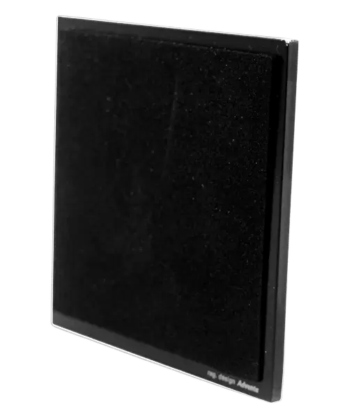
Glass Coaster
Individual Glass Coaster. Stylish and elegant polished safety glass, toughened and heat resistant (10x10cm, 7mm thick). Price shown is per individual coaster.
Individual Glass Coaster. Elegant polished safety toughened glass and heat resistant, matching Place Mats are also available
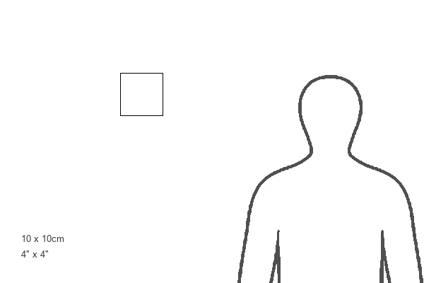
Estimated Image Size (if not cropped) is 7.6cm x 6.5cm (3" x 2.6")
Estimated Product Size is 10cm x 10cm (3.9" x 3.9")
These are individually made so all sizes are approximate
Artwork printed orientated as per the preview above, with landscape (horizontal) orientation to match the source image.
EDITORS COMMENTS
This print showcases a knee injury captured through a state-of-the-art 3D CT scan. The image reveals the intricate details of the knee joint, with a clear indication of a meniscal tear highlighted by arrows. Menisci, which are fibrocartilage crescents acting as shock absorbers between the femur and tibia, play a crucial role in maintaining joint health. The tear in the meniscus depicted here can result from trauma, often associated with sports injuries, or from gradual wear and tear over time. Treatment options vary depending on the severity of the tear; minor tears may respond well to physical therapy and anti-inflammatory medications while more severe cases necessitate surgical intervention. Radiating an air of medical expertise and precision diagnosis, this photograph offers valuable insights into understanding knee injuries. It emphasizes how critical it is to address such issues promptly for optimal recovery. With its vibrant colors highlighting areas of damage within this complex joint structure, this image serves as an educational tool for healthcare professionals and individuals seeking knowledge about knee injuries. By providing an up-close view of bones, cartilage, and damaged tissue in three dimensions using cutting-edge technology like CT scans, it allows us to delve deeper into our understanding of human anatomy and pathology. Captured by ZEPHYR/SCIENCE PHOTO LIBRARY's skilled photographers without any commercial intent mentioned in their caption notes; this print stands as both visually striking artwork and informative scientific documentation.
MADE IN THE UK
Safe Shipping with 30 Day Money Back Guarantee
FREE PERSONALISATION*
We are proud to offer a range of customisation features including Personalised Captions, Color Filters and Picture Zoom Tools
SECURE PAYMENTS
We happily accept a wide range of payment options so you can pay for the things you need in the way that is most convenient for you
* Options may vary by product and licensing agreement. Zoomed Pictures can be adjusted in the Basket.