
Fibrocartilage Collection
Fibrocartilage: Unseen Hero in Joint Health and Injury Recovery (Anatomy of Human Knee Joint) Fibrocartilage, a vital yet often overlooked component of our joints


All Professionally Made to Order for Quick Shipping
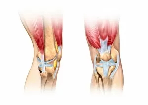
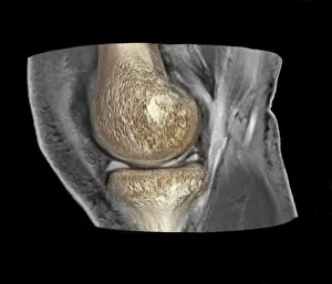
Fibrocartilage: Unseen Hero in Joint Health and Injury Recovery (Anatomy of Human Knee Joint) Fibrocartilage, a vital yet often overlooked component of our joints, plays a crucial role in maintaining their health and facilitating smooth movement. This connective tissue, visible in the human knee cutaway illustration, lines the joint surfaces and acts as a shock absorber, reducing friction and preventing wear. When injury strikes, as depicted in these 3D CT scans of knee injuries (C018 / 0648, C018 / 0639, C018 / 0645), fibrocartilage may sustain damage, leading to pain and impaired function. However, its remarkable ability to heal and regenerate makes it an essential focus in injury recovery. Fibrocartilage is not limited to the knee joint; it also protects the vertebral bodies in the spine, as seen in this artwork of the human backbone. Similarly, the hand bones and ligaments, as well as the lumbar spine and sacrum, benefit from the protective and cushioning properties (computer artwork). Understanding fibrocartilage's role in our anatomy is essential for appreciating its importance in overall health and injury recovery. Its resilience and regenerative properties make it a vital component in maintaining our mobility and quality of life.