Home > Popular Themes > Human Body
Lung, X-ray

![]()
Wall Art and Photo Gifts from Science Photo Library
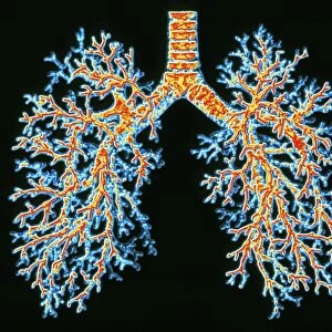
Lung, X-ray
Lung. Coloured bronchography (X-ray) of a healthy human lung. A contrast medium has been added to show the network of airways (green) in the right lung. The trachea (wind pipe) enters the lungs and splits into the two bronchi (one seen, upper right). The bronchi then further divide into bronchioles, which continue to divide and decrease in diameter. The bronchioles eventually end in groups of tiny air sacs known as alveoli (not seen). Alveoli are the site of gaseous exchange between the lungs and blood; oxygen enters the blood and carbon dioxide leaves
Science Photo Library features Science and Medical images including photos and illustrations
Media ID 6449863
© CNRI/SCIENCE PHOTO LIBRARY
Airway Airways Branches Branching Breathing Bronchi Bronchiole Bronchioles Bronchus Chest Contrast Medium False Colour Gas Exchange Gaseous Exchange Lung Net Work Pulmonary Radio Opaque Radiography Respiration Respiratory Ribs System Technique Thoracic Thorax Trachea X Ray Machine False Coloured
EDITORS COMMENTS
This print showcases a coloured bronchography X-ray of a healthy human lung, providing an intricate view of its internal structure. The image reveals the complex network of airways within the right lung, highlighted in vibrant green due to the addition of a contrast medium. At the center, we can observe the trachea or windpipe entering the lungs and dividing into two bronchi, with one visible in the upper right corner. As we delve deeper into this remarkable organ's anatomy, we witness further branching as the bronchi divide into smaller bronchioles. These bronchioles continue to decrease in diameter until they culminate in tiny air sacs called alveoli (not visible here). It is within these alveoli that gaseous exchange occurs between our lungs and blood vessels. Oxygen enters our bloodstream while carbon dioxide exits it. The radiograph also offers insight into how respiration functions within our bodies. Each breath we take relies on this intricate system of branching airways and delicate structures working harmoniously together. This stunning visual representation not only highlights the complexity and beauty of our respiratory system but also serves as a reminder of its vital role in sustaining life.
MADE IN THE UK
Safe Shipping with 30 Day Money Back Guarantee
FREE PERSONALISATION*
We are proud to offer a range of customisation features including Personalised Captions, Color Filters and Picture Zoom Tools
SECURE PAYMENTS
We happily accept a wide range of payment options so you can pay for the things you need in the way that is most convenient for you
* Options may vary by product and licensing agreement. Zoomed Pictures can be adjusted in the Basket.