
Trachea Collection
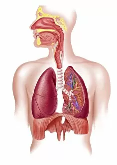
The trachea, also known as the windpipe, is a vital part of our respiratory system. It serves as a pathway for air to travel from the nose and mouth to the lungs


All Professionally Made to Order for Quick Shipping
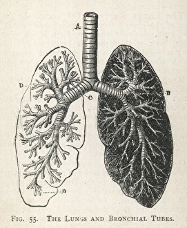
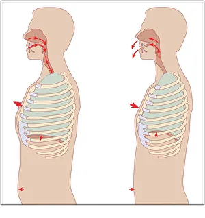
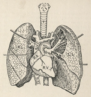
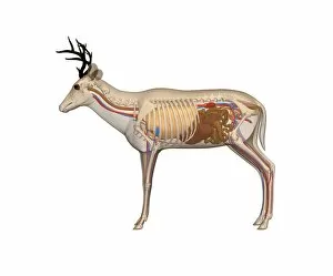
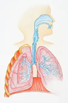
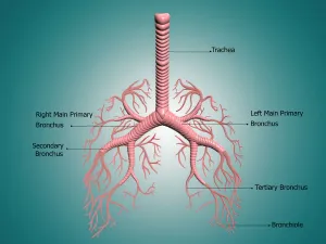
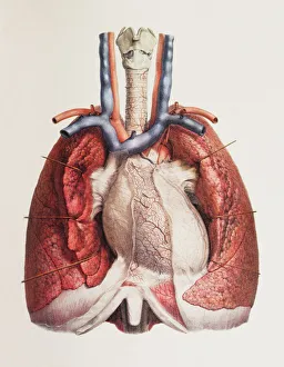
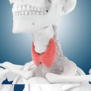
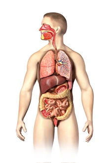
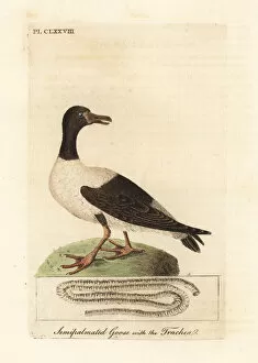
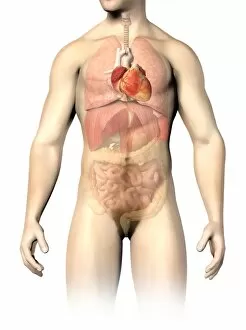
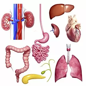
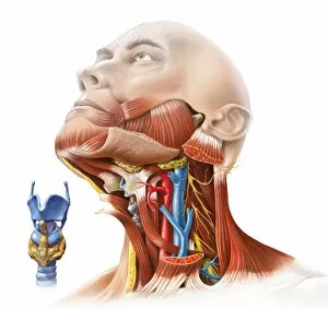
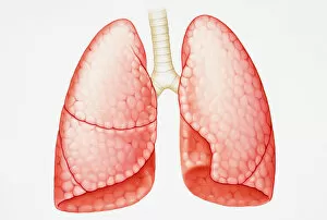
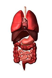
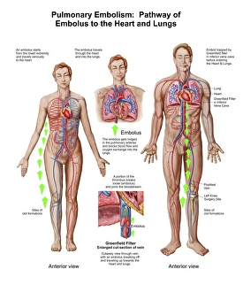
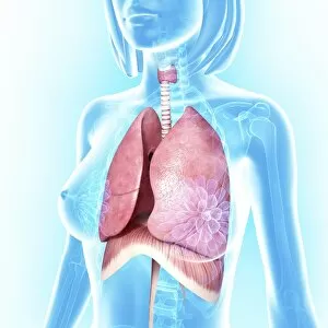
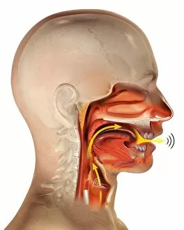
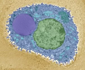
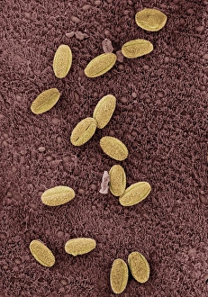
The trachea, also known as the windpipe, is a vital part of our respiratory system. It serves as a pathway for air to travel from the nose and mouth to the lungs. To understand its role better, let's take a look at some diagrams. In one diagram, we can see the intricate network of bronchial tubes that branch out from the trachea into both lungs. This branching structure ensures that oxygen reaches every corner of our lungs efficiently. Another diagram shows us how the heart, lungs, and windpipe are interconnected. The trachea sits right in front of the esophagus and behind the sternum bone, protecting it from any external pressure or damage. Understanding the mechanics of respiration is crucial when studying the trachea's function. A detailed diagram illustrates how inhalation and exhalation occur through this essential tube. Moving on to an illustration of our respiratory system, we can observe various components such as oral cavity, nasal cavity, larynx (voice box), trachea, bronchus (plural: bronchi), and lungs working together harmoniously to ensure proper breathing. Now let's focus on specific aspects related to the trachea itself. An anatomy depiction showcases details about bronchus and bronchial tubes – their structure and location within our body. Considering muscles' involvement in neck movements during respiration is important too; another image highlights these muscles' placement around our neck area. Zooming in further reveals fascinating microscopic views like scanning electron microscopy (SEM) images showing us close-ups of tracheal lining cells – giving us insights into their unique structures responsible for filtering air particles before they reach our delicate lung tissues. Exploring related anatomical features brings us artwork depicting thyroid anatomy since it lies just below where our windpipe splits into two main bronchi leading towards each lung. Let's not forget about other species.