
Bronchiole Collection
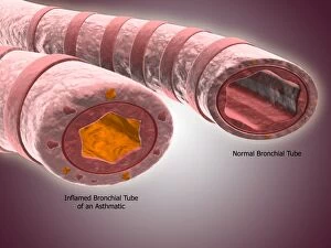
The bronchiole is a vital component of the respiratory system, specifically within the anatomy of the bronchus and bronchial tubes


All Professionally Made to Order for Quick Shipping
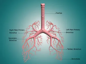
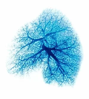
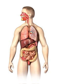
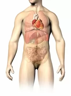
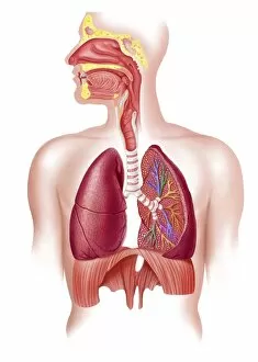
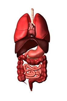
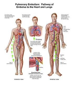
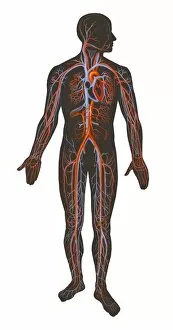
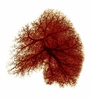
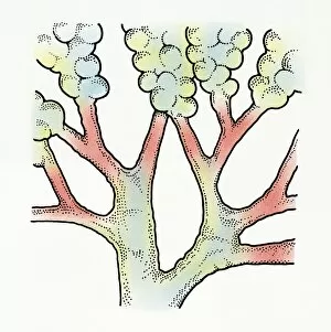
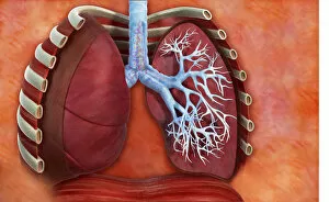
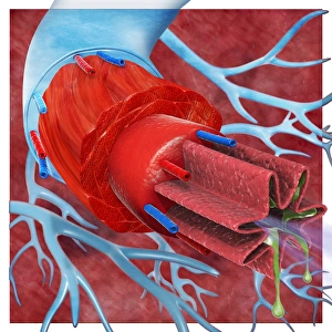
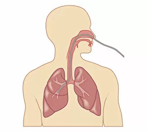
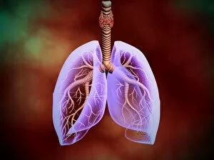
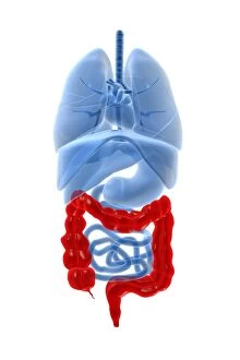
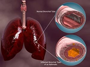
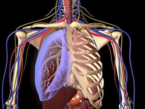
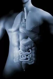
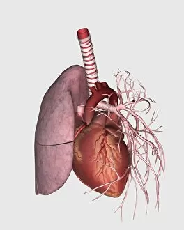
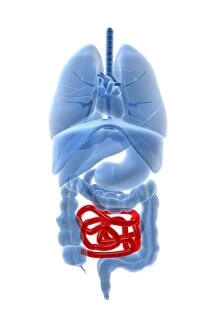
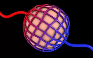
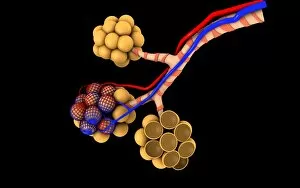
The bronchiole is a vital component of the respiratory system, specifically within the anatomy of the bronchus and bronchial tubes. These delicate structures are responsible for carrying air into and out of the lungs, ensuring proper oxygenation and removal of carbon dioxide. When examining an X-ray of the lung, one can observe these intricate passageways branching off from the main bronchi. They resemble a tree-like structure, with smaller branches known as bronchioles extending deeper into the lung tissue. In understanding male chest anatomy, it becomes evident that the they are nestled within the thorax alongside other essential organs such as the heart, veins, arteries, and lungs. This interconnectedness highlights their crucial role in maintaining respiratory function. Examining internal organ anatomy reveals how intricately woven together systems like respiration and circulation truly are. The human circulatory system relies on efficient oxygen exchange facilitated by healthy bronchioles to ensure optimal functioning throughout all bodily processes. A cutaway diagram provides a comprehensive view of how our respiratory system operates. It showcases how each individual lung is connected to its respective primary bronchus through numerous secondary and tertiary branches called bronchioles. Conceptual images further emphasize this interplay between different systems within our bodies. They illustrate how internal organs belonging to both respiratory and digestive systems coexist harmoniously while performing their distinct functions. Studying pulmonary embolism sheds light on potential complications that can arise within these pathways. An embolus traveling through veins may reach its final destination in either heart or lungs via various routes involving intricate networks of arteries and veins surrounding them. Understanding arterial-venous relationships across our body aids in comprehending blood flow dynamics associated with respiration. Arteries carry freshly oxygenated blood from lungs to tissues while veins return deoxygenated blood back for reoxygenation – all made possible by well-functioning bronchioles among other components involved in gas exchange.