
Respiration Collection
"Unlocking the Secrets of Respiration: From Lungs to Mitochondria" In this captivating journey through the intricate world of respiration


All Professionally Made to Order for Quick Shipping
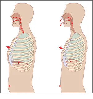
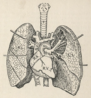
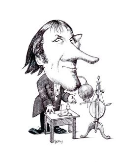
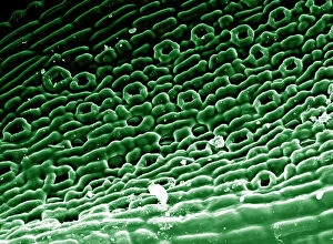
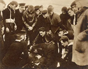
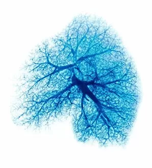
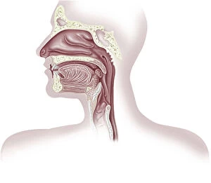
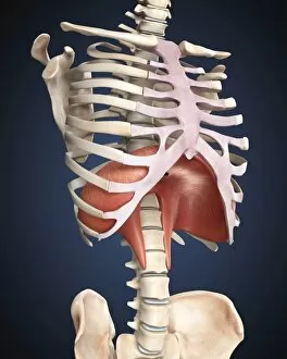
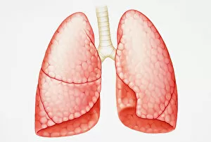
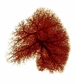
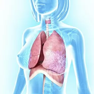
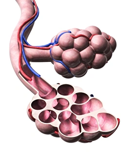
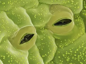
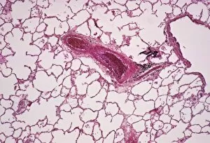
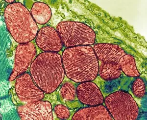
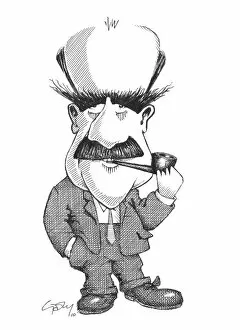
"Unlocking the Secrets of Respiration: From Lungs to Mitochondria" In this captivating journey through the intricate world of respiration, we explore the very essence of life itself. Starting with a detailed diagram showcasing the heart, lungs, and windpipe, we unravel the mechanics behind this vital process. Imagine a brave firefighter donning an oxygen mask amidst billowing smoke – a powerful reminder of how they are be a matter of life or death. Delving deeper into understanding its mechanisms, we encounter an enlightening diagram illustrating how each breath fuels our bodies' energy production within tiny powerhouses called mitochondria. Taking us back in time, an engraving from May 1872 reveals directions for restoring apparently dead individuals issued by the Royal Humane Society. This historical artifact serves as a testament to humanity's relentless pursuit to comprehend and preserve life through respiration. We also encounter Joseph Priestley's caricature - C015 / 6707 - paying homage to his groundbreaking discoveries on oxygen that paved the way for modern respiratory science. His contributions continue to inspire generations in their quest for knowledge about this fundamental process. A mesmerizing image - Picture No. 11675585 - showcases English oak leaf pores under scanning electron microscopy (SEM), highlighting nature's own version at work. These microscopic wonders remind us that every living organism relies on this essential exchange of gases for survival. However, not all stories surrounding it can filled with hope; cystic fibrosis emerges as a formidable challenge affecting countless lives worldwide. Yet even in adversity, remarkable figures like Konstantin Buteyko emerge – a Soviet doctor whose innovative breathing techniques have brought relief and improved quality of life for those battling respiratory conditions. Finally, our exploration concludes with another SEM image revealing trachea lining intricacies – yet another marvel that underscores nature's brilliance in designing efficient respiratory systems across species. Respiration is more than just inhaling and exhaling; it is the very essence of life.