Jigsaw Puzzle : Cornea, SEM


![]()
Jigsaw Puzzles from Science Photo Library
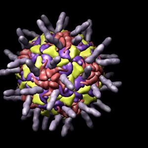
Cornea, SEM
Cornea. Coloured scanning electron micrograph (SEM) of a section through a cornea, the transparent region on the outer surface of the eye. The front of the eye is at top. The corneal epithelium (pink) lies above the substantia propria (parallel white/blue lines), the dense collagenous connective tissue that constitutes the bulk of the cornea. The cornea refracts (bends) light entering the eye onto the lens, thus helping to focus images onto the light-sensitive retina at the back of the eye. Magnification: x800 at 6x7cm size
Science Photo Library features Science and Medical images including photos and illustrations
Media ID 6449133
© STEVE GSCHMEISSNER/SCIENCE PHOTO LIBRARY
Connective Cornea Corneal Epithelial Epithelium Fibre Fibres Histological Histology Layer Layered Layers Parallel Sensory Sight Stratified Surface Tissue Vision Visual Sense Section Sectioned
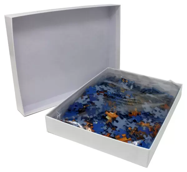
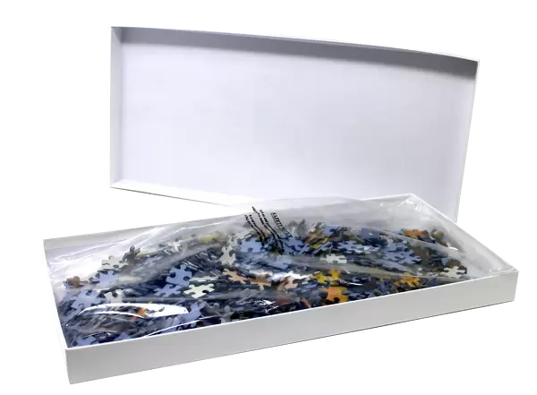
Jigsaw Puzzle (400 Pieces)
Discover the intricacies of the human body with our Media Storehouse Jigsaw Puzzles. This captivating puzzle features a coloured Scanning Electron Micrograph (SEM) image of a cornea section by Science Photo Library. Explore the intricacies of the cornea, the transparent outer layer of the eye, with this challenging and educational puzzle. Piece together the intricate details and bring the microscopic world to life in your living room. A perfect activity for families or individuals seeking a brain-teasing challenge and an opportunity to learn something new.
400 piece puzzles are custom made in the UK and hand-finished on 100% recycled 1.5 mm millboard. There is a level of repetition in jigsaw shapes with each matching piece away from its pair. The completed puzzle measures 31x47cm and is delivered packaged in an attractive presentation box specially designed to fit most letter box slots
Jigsaw Puzzles are an ideal gift for any occasion
Estimated Product Size is 47.2cm x 31.5cm (18.6" x 12.4")
These are individually made so all sizes are approximate
Artwork printed orientated as per the preview above, with landscape (horizontal) or portrait (vertical) orientation to match the source image.
EDITORS COMMENTS
This print showcases the intricate beauty of the cornea, the transparent region on the outer surface of our eyes. In this coloured scanning electron micrograph (SEM), we are granted a glimpse into the complex layers and structure that make up this vital part of our visual system. At first glance, we observe the front of the eye positioned at the top, with its attention-grabbing pink corneal epithelium resting above. Below it lies the substantia propria, an awe-inspiring dense collagenous connective tissue characterized by parallel white and blue lines. It is within this bulk of tissue that most of the cornea's composition resides. The significance of these layered structures becomes apparent as we learn about their role in vision. The cornea acts as a refractor, skillfully bending light rays that enter our eyes onto our lenses. This crucial function aids in focusing images onto our retina – a light-sensitive area located at the back of our eyes. With a magnification level set at x800 for a 6x7cm size print, every detail has been meticulously captured to provide us with an extraordinary view into this microscopic world. As we admire this image from Science Photo Library, let us marvel at how such complexity seamlessly combines to grant us one of life's greatest gifts – sight.
MADE IN THE UK
Safe Shipping with 30 Day Money Back Guarantee
FREE PERSONALISATION*
We are proud to offer a range of customisation features including Personalised Captions, Color Filters and Picture Zoom Tools
SECURE PAYMENTS
We happily accept a wide range of payment options so you can pay for the things you need in the way that is most convenient for you
* Options may vary by product and licensing agreement. Zoomed Pictures can be adjusted in the Basket.