Home > Science > SEM
Outer layers of the eye, SEM

![]()
Wall Art and Photo Gifts from Science Photo Library
Outer layers of the eye, SEM
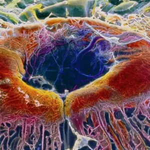
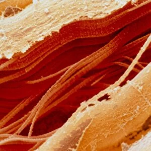
Outer layers of the eye. Coloured scanning electron micrograph (SEM) of the choroid (top) and sclera (bottom) layers of the eye. The sclera is the white outer layer of the eye. It is composed of tough fibrous connective tissue and covers all of the eye except the cornea. It protects the structures within the eyeball and attaches to muscles that move the eye and maintain its shape. The choroid layer, which lies behind the sclera and in front of the retina, is highly vascular and supplies blood to the back of the eye. It is pigmented to absorb excessive light, preventing internal reflections that would form multiple images on the retina. Magnification: x26 when printed 10 centimetres wide
Science Photo Library features Science and Medical images including photos and illustrations
Media ID 6448787
© STEVE GSCHMEISSNER/SCIENCE PHOTO LIBRARY
Brown Choroid Connective Tissue False Colour Fibrous Histological Histology Layer Layers Pigmented Protection Protective Sclera Sense Sight Vascular Vision False Coloured
FEATURES IN THESE COLLECTIONS
EDITORS COMMENTS
This print showcases the intricate outer layers of the eye, revealing its remarkable anatomy and protective capabilities. In this coloured scanning electron micrograph (SEM), we are presented with a close-up view of two crucial layers: the choroid and sclera. The sclera, depicted at the bottom, is responsible for safeguarding all structures within the eyeball except for the cornea. Composed of tough fibrous connective tissue, it acts as a shield against external harm while also supporting and maintaining the eye's shape. Its white appearance gives our eyes their characteristic look. Above it lies the highly vascularized choroid layer, which plays a vital role in nourishing and supplying blood to the back of our eyes. This pigmented layer not only absorbs excessive light but also prevents internal reflections that could lead to multiple images forming on our retina. Through this stunning SEM image, we gain an appreciation for both form and function within our visual system. The magnification used here allows us to observe these delicate details at 26 times their actual size when printed 10 centimetres wide. Science Photo Library has once again captured nature's wonders through their lens, reminding us of how truly extraordinary even seemingly ordinary parts of our body can be.
MADE IN THE UK
Safe Shipping with 30 Day Money Back Guarantee
FREE PERSONALISATION*
We are proud to offer a range of customisation features including Personalised Captions, Color Filters and Picture Zoom Tools
SECURE PAYMENTS
We happily accept a wide range of payment options so you can pay for the things you need in the way that is most convenient for you
* Options may vary by product and licensing agreement. Zoomed Pictures can be adjusted in the Basket.