
Connective Collection
"Exploring the Intricate Web of Connective Tissues: A Journey through SEM Images" Delving into the microscopic world


All Professionally Made to Order for Quick Shipping
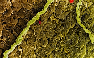
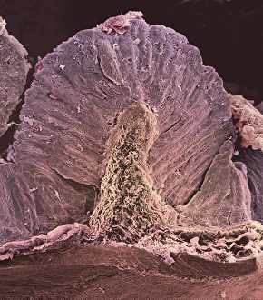
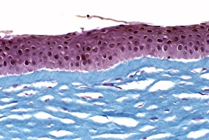
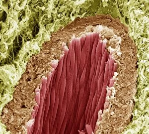
"Exploring the Intricate Web of Connective Tissues: A Journey through SEM Images" Delving into the microscopic world, we uncover a fascinating realm tissues that form an intricate network within our bodies. Through scanning electron microscopy (SEM), these images offer glimpses into the diverse structures and functions they serve. Starting with connective tissue fibers, we witness their remarkable arrangement resembling a finely woven tapestry. These resilient fibers provide strength and support to various organs and body parts, ensuring stability in our upper body anatomy. Moving on to fat cells captured under SEM, we observe their unique morphology. Some images reveal empty fat cells, reminding us of the dynamic nature of adipose tissue as it fluctuates in response to energy storage or release. Venturing deeper into our internal systems, SEM unveils captivating views of the intestinal lining. The delicate folds and microvilli present here play crucial roles in nutrient absorption and digestion processes within our stomach walls – another marvel showcased by SEM imagery. Shifting focus towards lactating breast tissue, SEM exposes its intricate architecture responsible for milk production during breastfeeding. This glimpse reminds us of the incredible capabilities of female reproductive organs like fallopian tubes – depicted through stunning SEM visuals that showcase their complex structure. As we explore this mesmerizing world hidden from plain sight, these captivating images remind us that beneath our skin lies an interconnected web facilitating vital bodily functions. From providing structural support to storing energy reserves or enabling reproduction processes – connective tissues truly embody the essence of unity within our biological framework.