Glass Frame : Fallopian tube


![]()
Mounted Prints from Science Photo Library
Fallopian tube
Fallopian tube. Coloured scanning electron micrograph (SEM) of the epithelium of a fallopian tube (oviduct), one of two muscular tubes connecting the ovary to the uterus (womb). There are two different types of epithelial columnar cells: ciliated (yellow, hair-like) and secretory (orange). The beat of the ciliated cells facilitates the transport of the ovum (egg) from the ovary towards the uterus. The secretory cells are covered by a vast number of microvilli and secrete substances that maintain a moist environment and provide nutrients to the ovum
Science Photo Library features Science and Medical images including photos and illustrations
Media ID 6450719
© SUSUMU NISHINAGA/SCIENCE PHOTO LIBRARY
Epithelium Fallopian Tube False Colour Microvilli Oviduct Re Production Reproductive System Secretory Cell Tissue Uterine Tube Cells False Coloured
8"x6" Glass Mount
Wall mounted or free-standing, these black edged glass frames feature a smooth chamfered edge and a stylish black border (on back face of the glass). Manufactured from 4mm thick glass, Glass Mounts are a durable, professional way of displaying and protecting your prints. Your 8x6 print is slotted into the back of the frame so can easily be changed if needed.
Tempered Glass Mounts are ideal for wall display, plus the smaller sizes can also be used free-standing via an integral stand
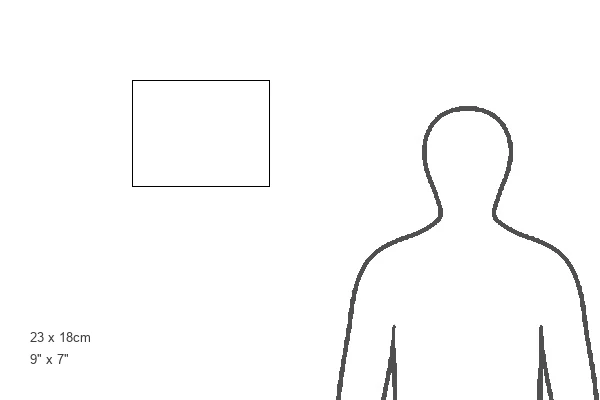
Estimated Image Size (if not cropped) is 20.3cm x 15.2cm (8" x 6")
Estimated Product Size is 22.8cm x 17.7cm (9" x 7")
These are individually made so all sizes are approximate
Artwork printed orientated as per the preview above, with landscape (horizontal) orientation to match the source image.
EDITORS COMMENTS
This print showcases the intricate beauty of a fallopian tube, captured through the lens of a scanning electron microscope. The fallopian tube, also known as an oviduct, is a vital component of the female reproductive system. It serves as one of two muscular tubes that connect the ovary to the uterus. The image reveals two distinct types of epithelial columnar cells within the fallopian tube: ciliated and secretory cells. The ciliated cells, depicted in vibrant yellow hues resembling delicate hair-like structures, play a crucial role in facilitating the transportation of eggs from the ovary towards the uterus. Their rhythmic beating propels these precious ova along their journey. Contrasting with their ciliated counterparts are secretory cells portrayed in striking orange tones. These specialized cells are adorned with countless microvilli and secrete substances that create a moist environment while providing essential nutrients for developing eggs. Through this stunning visual representation, we gain insight into both form and function within this complex biological system. This photograph not only highlights its scientific significance but also captivates viewers with its artistic appeal. Science Photo Library has once again succeeded in capturing nature's wonders at microscopic levels, offering us an awe-inspiring glimpse into our own anatomy and reproduction process.
MADE IN THE UK
Safe Shipping with 30 Day Money Back Guarantee
FREE PERSONALISATION*
We are proud to offer a range of customisation features including Personalised Captions, Color Filters and Picture Zoom Tools
SECURE PAYMENTS
We happily accept a wide range of payment options so you can pay for the things you need in the way that is most convenient for you
* Options may vary by product and licensing agreement. Zoomed Pictures can be adjusted in the Basket.