Glass Frame > Popular Themes > DNA
Glass Frame : Cell structure


![]()
Mounted Prints from Science Photo Library
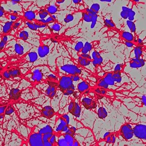
Cell structure
Cell structure. Confocal light micrograph of cultured endothelial cells. A fluorescent dye has been used to show the cell structure. Nuclei (blue) contain the cells genetic information, DNA (deoxyribonucleic acid) packaged in chromosomes. Actin filaments part of the cell cytoskeleton are green. The cytoskeleton is responsible for the structure and motility of the cell. Actin is the most abundant cellular protein. The golgi apparatus, or body (red), is a membrane-bound organelle that modifies and packages proteins. It consists of long flattened vesicles arranged in stacks. Endothelial cells are flat and line all of the bodys blood vessels
Science Photo Library features Science and Medical images including photos and illustrations
Media ID 6400349
© DAVID BECKER/SCIENCE PHOTO LIBRARY
Actin Apparatus Bodies Body C Ulture Cellular Confocal Light Micrograph Confocal Light Microscope Cultured Cytology Cytoskeleton Dyes Edothelial Endothelia Endothelium F Actin Filament Filamentous Filaments Fluorescence Fluorescent Fluorescent Dye Golgi Immunofluorescence Immunofluorescent Immunostained Membrane Bound Motility Nuclei Nucleus Organelle Stained Structural Vascular Cells Protein
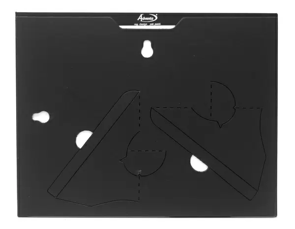
8"x6" Glass Mount
Wall mounted or free-standing, these black edged glass frames feature a smooth chamfered edge and a stylish black border (on back face of the glass). Manufactured from 4mm thick glass, Glass Mounts are a durable, professional way of displaying and protecting your prints. Your 8x6 print is slotted into the back of the frame so can easily be changed if needed.
Tempered Glass Mounts are ideal for wall display, plus the smaller sizes can also be used free-standing via an integral stand
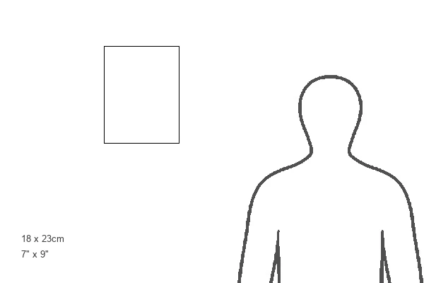
Estimated Image Size (if not cropped) is 15.2cm x 20.3cm (6" x 8")
Estimated Product Size is 17.7cm x 22.8cm (7" x 9")
These are individually made so all sizes are approximate
Artwork printed orientated as per the preview above, with portrait (vertical) orientation to match the source image.
EDITORS COMMENTS
This print showcases the intricate structure of endothelial cells, as captured through a confocal light microscope. The fluorescent dye used in this image highlights various components of the cell, revealing its inner workings. The blue nuclei contain DNA, which carries the cell's genetic information and is packaged in chromosomes. Green actin filaments form part of the cell cytoskeleton, responsible for maintaining its structure and enabling movement. Actin is a vital protein found abundantly within cells. The red-colored golgi apparatus stands out prominently in this image; it is a membrane-bound organelle that plays a crucial role in modifying and packaging proteins within the cell. Composed of long flattened vesicles arranged in stacks, it ensures efficient protein transport. Endothelial cells are flat and line all blood vessels throughout our bodies. They play an essential role in vascular biology by regulating blood flow and vessel permeability. This stunning micrograph not only provides valuable insights into cellular structures but also serves as a testament to the remarkable complexity present within even seemingly simple organisms like individual cells. It reminds us of the beauty hidden beneath our skin – an intricate world that fuels life itself.
MADE IN THE UK
Safe Shipping with 30 Day Money Back Guarantee
FREE PERSONALISATION*
We are proud to offer a range of customisation features including Personalised Captions, Color Filters and Picture Zoom Tools
SECURE PAYMENTS
We happily accept a wide range of payment options so you can pay for the things you need in the way that is most convenient for you
* Options may vary by product and licensing agreement. Zoomed Pictures can be adjusted in the Basket.