
Vascular Collection
"Exploring the Intricate World Anatomy: From Brain Blood Vessels to Maize Roots" Delve into the fascinating realm anatomy


All Professionally Made to Order for Quick Shipping
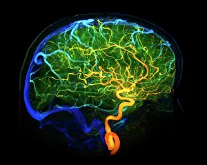
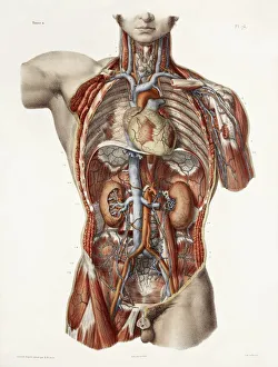
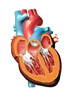
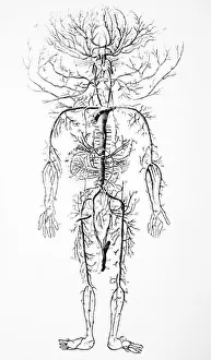
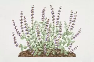
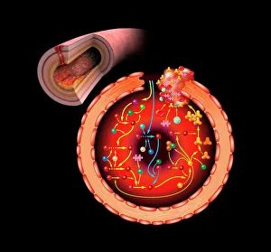
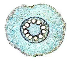
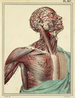
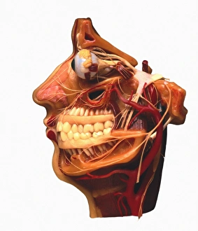
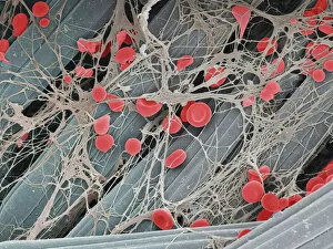
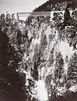
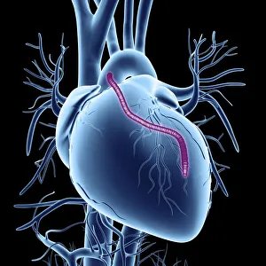
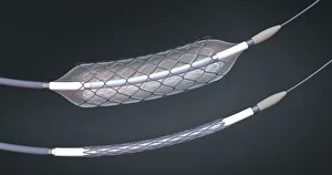
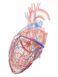
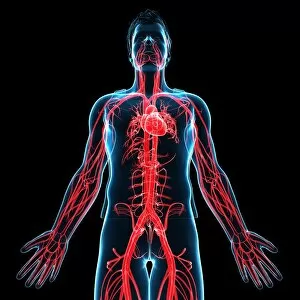
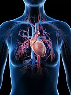
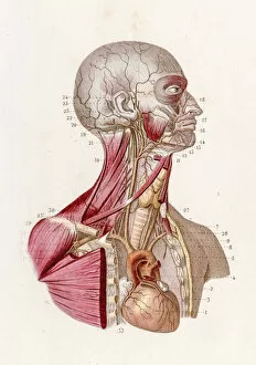
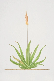
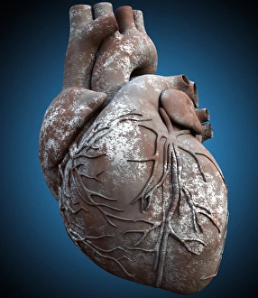
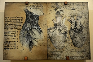
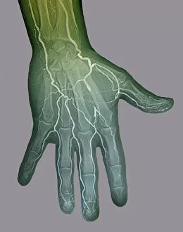
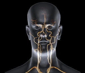
"Exploring the Intricate World Anatomy: From Brain Blood Vessels to Maize Roots" Delve into the fascinating realm anatomy, where a complex network of blood vessels ensures our bodies function seamlessly. Witness the marvels of this intricate system through various historical artworks and scientific imagery. Travel back in time to 1981 with a mesmerizing 3D angiogram C007, showcasing the intricate web of brain blood vessels. Marvel at how these delicate pathways nourish our most vital organ, ensuring its optimal performance. Step into the world of cardiovascular system exploration as you uncover historical artwork depicting its complexity. Admire exquisite neck anatomy illustrations from the 19th century, capturing every detail with artistic precision. Discover the hidden beauty within human heart anatomy through stunning artwork that showcases its awe-inspiring structure. Dive deep into cell structures that make up this remarkable organ, unraveling its secrets one layer at a time. Journey through time as you explore arterial systems depicted in captivating 18th-century artwork. Be captivated by their elegance and understand their crucial role in maintaining overall health and vitality. Immerse yourself in neck vascular anatomy captured by historical artists who sought to document this enigmatic region's intricacies. Let your eyes trace each artery's path as it weaves through muscles and tissues. Witness X-ray images revealing neck and shoulder arteries' hidden mysteries—an opportunity to glimpse beneath our skin's surface and appreciate nature's design flawlessly crafted for optimum functionality. Marvel at Salvia officinalis or Common Sage plant—a botanical wonder known for promoting healthy circulation—its vibrant leaves symbolizing lifeblood coursing through our veins like an eternal river. Transport yourself back to 1825 when male groin arteries were meticulously illustrated—an homage to medical pioneers who unraveled body secrets long before modern technology existed. Uncover artistry behind blood coagulation cascade depicted in breathtaking artwork C016 / 9873.