Fraud Blocker Skip to main content

Nuclei Collection

"Nuclei: The Intricate Powerhouses within Our Cells and Beyond" Delving into the microscopic world, we encounter the mesmerizing beauty of nuclei

Background imageNuclei Collection: Cerebellum tissue, light micrograph

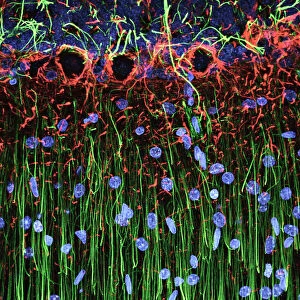
Cerebellum tissue, light micrograph
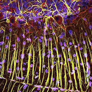
Cerebellum tissue. Confocal light micrograph of a section through the cerebellum of the brain. Purkinje cells, a type of neuron (nerve cell), are red
Background imageNuclei Collection: ATLAS detector, CERN

ATLAS detector, CERN
ATLAS detector. Engineer standing on a platform in front of the ATLAS (a torodial LHC apparatus) detector (circular) at CERN (the European particle physics laboratory) near Geneva, Switzerland

Background imageNuclei Collection: ATLAS detector, CERN

ATLAS detector, CERN
ATLAS detector. Composite image of the ATLAS (a torodial LHC apparatus) detector (circular) at CERN (the European particle physics laboratory) near Geneva, Switzerland

Background imageNuclei Collection: CMS detector, CERN

CMS detector, CERN
CMS detector. Part of the CMS (compact muon solenoid) detector at CERN (the European particle physics laboratory) near Geneva, Switzerland

Background imageNuclei Collection: Hippocampus brain tissue

Hippocampus brain tissue
Hippocampus tissue. Light micrograph of a sagittal (side view) section through the hippocampus of the brain showing the nerve cells within it

Background imageNuclei Collection: HeLa cells, light micrograph C017 / 8299

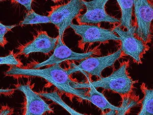
HeLa cells, light micrograph C017 / 8299
HeLa cells, multiphoton fluorescence micrograph (MFM). The cell nuclei, which contain the cells genetic information, are purple. Microtubules are blue and actin microfilaments are red

Background imageNuclei Collection: Brain tissue blood supply

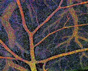
Brain tissue blood supply. Light micrograph of a section through cortex tissue from a brain, showing the blood vessels (branching) that supply it

Background imageNuclei Collection: Medulla oblongata in the brain, artwork

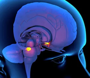
Medulla oblongata in the brain, artwork
Hypothalamus in the brain. Computer artwork of a persons head showing the left hemisphere of the brain inside. The highlighted area shows the hypothalamus

Background imageNuclei Collection: Nuclear Fission Artwork

Nuclear Fission Artwork
Nuclear fission. Conceptual computer artwork of an atom being split through nuclear, or atomic, fission (splitting). Electrons (orange) can be seen orbiting the nucleus (centre)

Background imageNuclei Collection: Kidney tubules in section

Kidney tubules in section
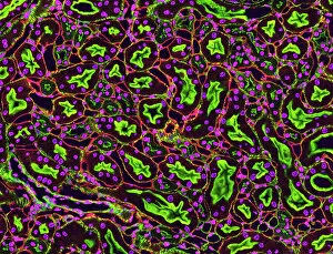
Kidney tubules. Fluorescent light micrograph of a section through kidney tissue showing numerous tubules (black/green). The tubules are seen in cross-section

Background imageNuclei Collection: Glial stem cell culture, light micrograph

Glial stem cell culture, light micrograph
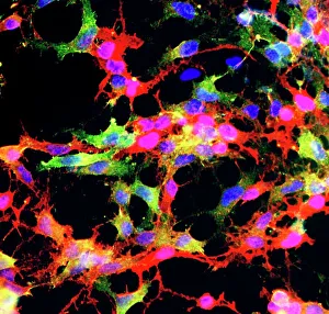
Glial stem cell culture. Fluorescent light micrograph of glial stem cells producing the protein NG2 (red) as they mature. These stem cells can differentiate into several types of glial cells

Background imageNuclei Collection: Cerebral cortex nerve cells

Cerebral cortex nerve cells. Confocal light micrograph of neurons (nerve cells, red) and glial cells (support cells, gold) from the cerebral cortex

Background imageNuclei Collection: Cell structure

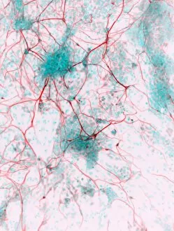
Cell structure. Confocal light micrograph of cultured endothelial cells. A fluorescent dye has been used to show the cell structure

Background imageNuclei Collection: HeLa cells, light micrograph C017 / 8298

HeLa cells, light micrograph C017 / 8298
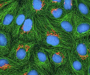
HeLa cells, multiphoton fluorescence micrograph (MFM). The cell nuclei, which contain the cells genetic information, are blue. Golgi bodies, which modify and package proteins, are orange

Background imageNuclei Collection: Mitosis, light micrograph

Mitosis, light micrograph
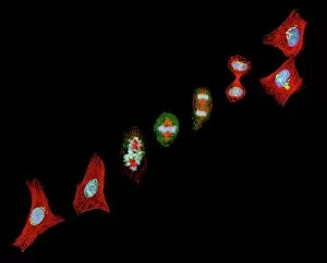
Mitosis. Confocal light micrograph of the stages of mitosis (nuclear division) and cytokinesis (cell division). During mitosis the nuclear envelope disintegrates (3rd image) and the chromosomes (blue)

Background imageNuclei Collection: Cerebellum tissue, light micrograph

Cerebellum tissue, light micrograph
Cerebellum tissue. Confocal light micrograph of a section through the cerebellum of the brain showing two types of glial cells (support cells); astrocytes (star-shaped)

Background imageNuclei Collection: Cerebellum tissue, light micrograph

Cerebellum tissue, light micrograph
Cerebellum tissue. Confocal light micrograph of a section through the cerebellum of the brain. Purkinje cells, a type of neuron (nerve cell), are red

Background imageNuclei Collection: Oligodendrocyte nerve cells

Oligodendrocyte nerve cells. Fluorescent light micrograph of human oligodendrocyte nerve cells. Cell nuclei, which contain the cells genetic information, have been dyed blue

Background imageNuclei Collection: Evolution of the Universe, artwork

Evolution of the Universe, artwork
Evolution of the Universe. Computer artwork showing the evolution of the Universe from the Big Bang (far left) 12-15 billion years ago to the present day (far right)

Background imageNuclei Collection: Astrocyte nerve cell

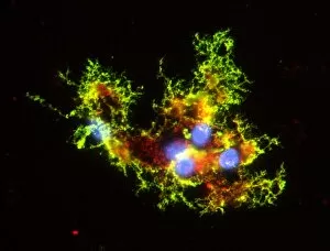
Astrocyte nerve cell. Fluorescent light micrograph of an astrocyte cell from a human brain. Intermediate filaments (IFs), part of the cells cytoskeleton, have been dyed green

Background imageNuclei Collection: Lymphocyte white blood cells, artwork

Lymphocyte white blood cells, artwork
Lymphocyte white blood cells. Computer artwork of lymphocyte white blood cells, showing their nuclei (red, centre). Lymphocytes, like all white blood cells, are part of the immune system

Background imageNuclei Collection: Heart muscle, confocal light micrograph

Heart muscle, confocal light micrograph
Heart muscle. Confocal light micrograph of a section through cardiac (heart) muscle. Cardiac muscle consists of branching elongated muscle cells

Background imageNuclei Collection: Antennae colliding galaxies C017 / 3745

Antennae colliding galaxies C017 / 3745
Antennae colliding galaxies, combined optical image. The Antennae (NGC 4038 and NGC 4039) are formed of two galaxies colliding due to mutual gravitational attraction

Background imageNuclei Collection: Picture No. 10874514

Picture No. 10874514
Light Micrograph (LM): A light micrograph of the nuclei of plant cells Date:

Background imageNuclei Collection: Stochastic gene expression, illustration C018 / 0906

Stochastic gene expression, illustration C018 / 0906
Stochastic gene expression, illustration. Every cell in an organism contains every single gene that makes up the organisms genome. However, they are not all active (expressed) in each cell

Background imageNuclei Collection: Artwork of nuclear fusion reaction

Artwork of nuclear fusion reaction
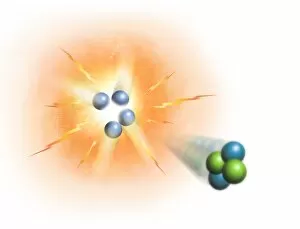
Conceptual image representing the process of nuclear fusion, specifically the creation of helium from hydrogen. Four protons (hydrogen nuclei) are combining on the left

Background imageNuclei Collection: Neural progenitor cell differentiation C018 / 8758

Neural progenitor cell differentiation C018 / 8758
Neural progenitor cell differentiation. Fluorescence light micrograph of neural progenitor cells that have been grown in a medium that is selective for astrocytes for three weeks

Background imageNuclei Collection: Myelin sheats and glial cells, artwork C014 / 2646

Myelin sheats and glial cells, artwork C014 / 2646
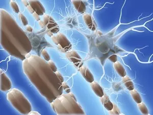
Computer artwork depicting axons surrounded by a myelin sheath (brown) and microglial cells (light blue). Glial cells are nervous system cells that provide structural support

Background imageNuclei Collection: Stem cell-derived retinal cells

Stem cell-derived retinal cells. Fluorescence light micrograph of retinal pigment epithelium (RPE) cells that have been derived from human embryonic stem cells (HESC)

Background imageNuclei Collection: Stem cell-derived neurons, micrograph

Stem cell-derived neurons, micrograph
Stem cell-derived neurons. Light micrograph of human nerve cells (neurons) that have been derived from induced pluripotent stem cells (IPS). Tuj1 proteins are cyan, and cell nuclei are red

Background imageNuclei Collection: Astrocyte brain cells, light micrograph

Astrocyte brain cells, light micrograph
Astrocyte brain cells. Fluorescence light micrograph of primary astrocyte cells from the brain of a mouse. Astrocytes have numerous branches of connective tissue that provide support

Background imageNuclei Collection: MDCK cells, inverted light micrograph C017 / 7647

MDCK cells, inverted light micrograph C017 / 7647
MDCK cells. Differential interference contrast inverted light micrograph of Madin-Darby canine kidney (MDCK) cells. This is a kidney epithelial cell line from a cocker spaniel

Background imageNuclei Collection: Cardiac muscle, light micrograph

Cardiac muscle, light micrograph
Cardiac muscle. Light micrograph of a section through muscle tissue from a heart, showing the cardiomyocyte cells. Heart muscle cells show central nuclei but the dominant feature is the abundance of

Background imageNuclei Collection: Peripheral nerve, light micrograph

Peripheral nerve, light micrograph
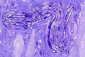
Peripheral nerve. Light micrograph of a section through a peripheral nerve. This is a mixed nerve with myelinated axons (dark blue circles)

Background imageNuclei Collection: Nerve ganglion, light micrograph

Nerve ganglion, light micrograph
Nerve ganglion. Light micrograph of a section through a dorsal (sensory) spinal root ganglion associated with a sensory nerve root of the spinal cord. Sensory information from peripheral sites e.g

Background imageNuclei Collection: Myelinated nerve, light micrograph

Myelinated nerve, light micrograph
Myelinated nerve. Light micrograph of a section through a peripheral myelinated nerve, showing many individual axons each covered with deep blue-staining myelin sheaths

Background imageNuclei Collection: Ductal breast cancer, TEM

Ductal breast cancer, TEM
Ductal breast cancer. Transmission electron micrograph (TEM) of a section through breast tissue, showing malignant (cancerous) epithelial cells arising from the milk ducts of the breast

Background imageNuclei Collection: Skin anatomy, artwork

Skin anatomy, artwork
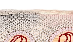
Skin anatomy. Artwork of a section through human skin, showing the flattening of skin cells as they rise to the dead keratinised surface layer (top) from the living layer below (the dermis)

Background imageNuclei Collection: Smooth muscle tissue, TEM

Smooth muscle tissue, TEM
Smooth muscle tissue. Transmission electron micrograph (TEM) of a longitudinal section through smooth muscle cells. Smooth muscle cells are spindle-shaped and often closely packed together

Background imageNuclei Collection: Heart muscle, light micrograph C016 / 0517

Heart muscle, light micrograph C016 / 0517
Heart muscle. Light micrograph of a section through heart (cardiac) muscle tissue, showing cardiomyocyte muscle cells. These cells have a centrally located nucleus and are branched

Background imageNuclei Collection: Nerve ganglion, light micrograph C016 / 0532

Nerve ganglion, light micrograph C016 / 0532
Nerve ganglion. Light micrograph of a section through a nerve ganglion of the peripheral nervous system, showing clusters of nerve cell bodies

Background imageNuclei Collection: Myelinated nerve fibres, light micrograph C016 / 0531

Myelinated nerve fibres, light micrograph C016 / 0531
Myelinated nerve fibres. Light micrograph of a section through a spinal sensory nerve containing many closely packed axons exhibiting a wave-type appearance

Background imageNuclei Collection: Myelin sheats and glial cells, artwork C014 / 2647

Myelin sheats and glial cells, artwork C014 / 2647
Computer artwork depicting axons surrounded by a myelin sheath (brown) and microglial cells (light blue). Glial cells are nervous system cells that provide structural support

Background imageNuclei Collection: Stem cell-derived nerve cells

Stem cell-derived nerve cells. Fluorescence light micrograph of neural (nerve) stem cells that have been derived from human embryonic stem cells (HESC)

Background imageNuclei Collection: Kidney tissue, confocal micrograph C014 / 4611

Kidney tissue, confocal micrograph C014 / 4611
Kidney tissue. Laser scanning confocal micrograph (LSCM) of a section through a human kidney, showing the cell nuclei (dark)

Background imageNuclei Collection: Liver portal triad, light micrograph C016 / 8490

Liver portal triad, light micrograph C016 / 8490
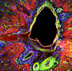
Liver portal triad. Fluorescence deconvolution micrograph of a section through a portal triad in liver tissue, showing hepatocyte cells (red)

Background imageNuclei Collection: Liver portal triad, light micrograph C016 / 8489

Liver portal triad, light micrograph C016 / 8489
Liver portal triad. Fluorescence deconvolution micrograph of a section through a portal triad in liver tissue, showing hepatocyte cells (red)

Background imageNuclei Collection: Liver portal triad, light micrograph C016 / 8488

Liver portal triad, light micrograph C016 / 8488
Liver portal triad. Fluorescence deconvolution micrograph of a section through a portal triad in liver tissue, showing hepatocyte cells (red)


All Professionally Made to Order for Quick Shipping

"Nuclei: The Intricate Powerhouses within Our Cells and Beyond" Delving into the microscopic world, we encounter the mesmerizing beauty of nuclei. In cerebellum tissue, a light micrograph reveals these vital command centers orchestrating our every move. Venturing deeper into scientific frontiers, we find ourselves at CERN's ATLAS detector, where nuclei play a crucial role in unraveling the mysteries of particle physics. Similarly, the CMS detector at CERN unveils their significance in understanding fundamental particles and forces. Shifting our focus to brain anatomy, hippocampus tissue showcases intricate nuclei that contribute to memory formation and spatial navigation. Meanwhile, HeLa cells captured under a light microscope exhibit their own unique nuclei patterns (C017 / 8299), highlighting their importance in medical research. Art meets science as an artwork depicting medulla oblongata reminds us of its critical role in regulating essential bodily functions through its specialized nuclei arrangement. Nuclear fission artwork further emphasizes how they are release immense energy when harnessed correctly. Zooming out to kidney tubules sectioned under a microscope slide unravels the presence of numerous cell nuclei responsible for maintaining fluid balance and waste elimination within our bodies. Expanding beyond traditional boundaries, glial stem cell culture offers insights into regenerative medicine with its vibrant display of proliferating nuclei (light micrograph). These versatile structures are central to cellular growth and differentiation processes. Examining cell structure more broadly uncovers how each nucleus houses genetic material that directs cellular activities like protein synthesis and DNA replication, and is this blueprint that shapes life itself. Lastly, exploring brain tissue blood supply highlights how oxygen-rich blood nourishes countless neuronal networks residing within diverse nuclear ensembles (HeLa cells - C017 / 8298). Intricate yet awe-inspiring, these glimpses into various realms remind us of the indispensable roles played by nucleic entities – from individual cells to complex systems – in shaping our understanding of life, physics, and medicine.