Fraud Blocker Skip to main content

Nuclei Collection (page 5)

"Nuclei: The Intricate Powerhouses within Our Cells and Beyond" Delving into the microscopic world, we encounter the mesmerizing beauty of nuclei

Background imageNuclei Collection: Fallopian tube, TEM

Fallopian tube, TEM
Fallopian tube, coloured transmission electron micrograph (TEM). Section through non-ciliated columnar epithelium from a fallopian tube. The ciliated cells are not shown here

Background imageNuclei Collection: Trachea mucous membrane, SEM

Trachea mucous membrane, SEM
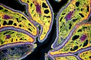
Trachea mucous membrane. Coloured scanning electron micrograph (SEM) of a fractured mucous membrane of the trachea (wind pipe), showing the epithelium and underlying connective tissue

Background imageNuclei Collection: Cerebellum structure, light micrograph

Cerebellum structure, light micrograph
Cerebellum structure. Coloured light micrograph of a section through the highly-folded cerebellum of the brain. The cerebellum comprises three main layers

Background imageNuclei Collection: Frogspawn, SEM

Frogspawn, SEM
Frogspawn. Coloured scanning electron micrograph (SEM) of a section through frog eggs, showing the large nucleus containing nucleoli, surrounded by yolk

Background imageNuclei Collection: Pancreas tissue, SEM

Pancreas tissue, SEM
Pancreas tissue. Coloured scanning electron micrograph (SEM) of fractured pancreas tissue, showing numerous acinar cells, containing secretory zymogen granules

Background imageNuclei Collection: Monocyte white blood cells, artwork

Monocyte white blood cells, artwork
Monocyte white blood cells. Computer artwork of monocyte white blood cells, showing their lobed nuclei (purple, centre). Monocytes, like all white blood cells

Background imageNuclei Collection: Basophil white blood cells, artwork

Basophil white blood cells, artwork
Basophil white blood cells. Computer artwork of basophil white blood cells, showing their lobed nuclei (red). Basophils are the smallest and least common of the white blood cells

Background imageNuclei Collection: Eosinophil white blood cells, artwork

Eosinophil white blood cells, artwork
Eosinophil white blood cells. Computer artwork of eosinophil white blood cells, showing their lobed nuclei (centre). Eosinophils, like all white blood cells, are part of the immune system

Background imageNuclei Collection: Cytokinesis, artwork

Cytokinesis, artwork
Cytokinesis. Artwork showing the stage of cell division that involves the splitting of the cell cytoplasm between two daughter cells

Background imageNuclei Collection: Cell division, artwork

Cell division, artwork
Cell division. Computer artwork of an animal cell undergoing mitosis (nuclear division) and cytokinesis (cell division) to form two identical daughter cells (bottom)

Background imageNuclei Collection: Cytokinesis, diagram

Cytokinesis, diagram
Cytokinesis. Diagram showing the stage of cell division that involves the splitting of the cell cytoplasm between two daughter cells

Background imageNuclei Collection: Strychnine seed tissue, light micrograph

Strychnine seed tissue, light micrograph
Strychnine seed tissue. Light micrograph of a section through a seed of the strychnine plant (Strychnos nux vomica), showing the cellular bridges (plasmodesma)

Background imageNuclei Collection: Cell manipulation

Cell manipulation. Cells being manipulated with laboratory instruments as part of research into cell biology. The dark areas are the cell nuclei

Background imageNuclei Collection: Kidney cells, light micrograph

Kidney cells, light micrograph
Kidney cells. Quantum dot fluorescence micrograph of a section through kidney tissue showing its cells

Background imageNuclei Collection: Embryonic stem cells, light micrograph

Embryonic stem cells, light micrograph
Embryonic stem cells. Fluorescence light micrograph of human embryonic neural stem cells forming neuronal networks. Tubulin protein is red; cell nuclei are blue

Background imageNuclei Collection: Adipose stem cells, light micrograph

Adipose stem cells, light micrograph
Adipose stem cells. Fluorescence light micrograph of human stem cells derived from adipose (fat) tissue. Nestin protein filaments are red; membrane cofactor protein is green;

Background imageNuclei Collection: Lung cells, fluorescent micrograph

Lung cells, fluorescent micrograph
Lung cells. Immunofluorescence light micrograph of pulmonary endothelial cells. Endothelial cells are specialized epithelial cells that line the inner surface of blood vessels

Background imageNuclei Collection: DNA packaging, artwork

DNA packaging, artwork
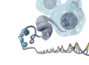
DNA packaging. Computer artwork showing how DNA (deoxyribonucleic acid) is packaged within cells. Two DNA strands, consisting of a sugar-phosphate backbone attached to nucleotide bases

Background imageNuclei Collection: Keratinocyte skin cells, light micrograph

Keratinocyte skin cells, light micrograph
Keratinocyte skin cells. Fluorescent light micrograph of the cytoskeleton of human keratinocyte skin cells. Cell nuclei are oval. The rest of the cells contents have been biochemically extracted

Background imageNuclei Collection: Macrophage cells, TEM

Macrophage cells, TEM
Macrophage cells, coloured transmission electron micrograph (TEM). The cell nuclei are purple. Mitochondria (dark pink ovals) in the cytoplasm produce energy for the cell

Background imageNuclei Collection: Simulating radioactive decay

Simulating radioactive decay. Small cubes used to simulate the random action of radioactive decay. Each block represents a nucleus of a radioactive material. Each has one black side

Background imageNuclei Collection: Cartilage cells, TEM

Cartilage cells, TEM
Cartilage cells. Coloured transmission electron micrograph (TEM) of a section through chondrocytes from nasal hyaline cartilage

Background imageNuclei Collection: Bird red blood cells, SEM

Bird red blood cells, SEM
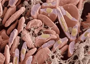
Bird red blood cells. Coloured scanning electron micrograph (SEM) of fractured red blood cells (erythrocytes, red) from avian heart tissue

Background imageNuclei Collection: Cellular packaging of DNA, artwork

Cellular packaging of DNA, artwork
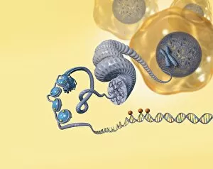
Cellular packaging of DNA. Artwork of a strand of the genetic material DNA (deoxyribonucleic acid) unwound from the nucleus (blue) of a cell (orange, upper right)

Background imageNuclei Collection: Virus particles entering cells, artwork

Virus particles entering cells, artwork
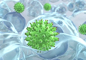
Virus particles (green) entering cells, computer artwork. The cell nuclei (dark blue) are also seen. The spikes on the virus particles are surface proteins that help them to attach to the cell

Background imageNuclei Collection: Neural stem cells in culture

Neural stem cells in culture
Neural stem cell in culture, fluorescent light micrograph. The stem cells have been dyed for nestin (red), an intermediate filament (IF) protein, and the nuclei are dyed blue

Background imageNuclei Collection: Brain cells in culture, light micrograph

Brain cells in culture, light micrograph
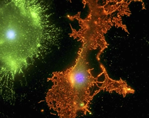
Brain cells in culture. Fluorescent light micrograph of a microglial cell (upper left) and an oligodendrocyte (centre) from a human brain

Background imageNuclei Collection: Reptilian red blood cells, SEM

Reptilian red blood cells, SEM
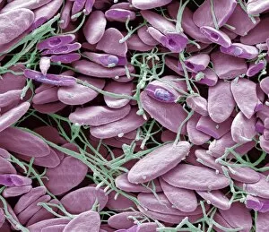
Reptilian red blood cells. Coloured scanning electron micrograph (SEM) of whole and fractured red blood cells (erythrocytes, purple) from reptilian tissue

Background imageNuclei Collection: Spirogyra algae, light micrograph

Spirogyra algae, light micrograph
Spirogyra algae. Light micrograph of filaments from a Spirogyra sp. alga, showing its spiralling bands of chloroplasts (green). Central nuclei (round) are also seen in each cell

Background imageNuclei Collection: Stomatal complex, TEM

Stomatal complex, TEM
Stomal complex. Coloured transmission electron micrograph (TEM) of a stomatal (pore) complex in the young leaf of the pea plant (Pisum sativum)

Background imageNuclei Collection: Poppy ovary with developing seeds, LM

Poppy ovary with developing seeds, LM
Poppy ovary with developing seeds. Light micrograph (LM) of a cross-section through the centre of a mature poppy ovary, showing placentas bearing developing seeds (red)

Background imageNuclei Collection: Mature poppy ovary, light micrograph

Mature poppy ovary, light micrograph
Mature poppy ovary. Light micrograph (LM) of a cross-section through a mature poppy ovary, which is incompletely divided by septa (membranes) that do not meet in the centre

Background imageNuclei Collection: Atom, artwork

Atom, artwork
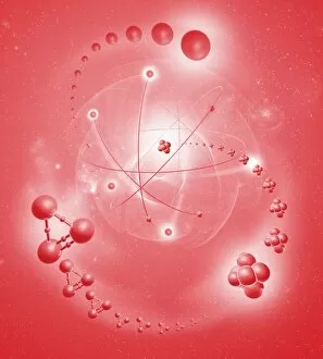
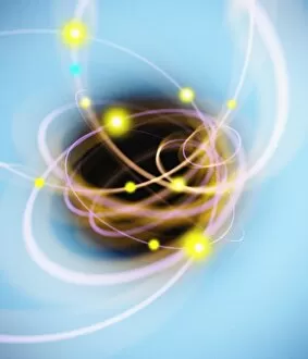
Atomic structure. Conceptual computer artwork of nine electrons orbiting a central nucleus. Other particles are seen around the atom. This is a classical schematic Bohr model of an atom

Background imageNuclei Collection: Subatomic particles abstract

Subatomic particles abstract

Background imageNuclei Collection: Intestinal smooth muscle cells, TEM

Intestinal smooth muscle cells, TEM
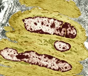
Intestinal smooth muscle cells. Coloured transmission electron micrograph (TEM) of a section through several smooth muscle cells from the intestines

Background imageNuclei Collection: Thyroid follicle structures, TEM

Thyroid follicle structures, TEM
Thyroid follicle structures. Coloured transmission electron micrograph (TEM) of a section through structures in a thyroid follicle


All Professionally Made to Order for Quick Shipping

"Nuclei: The Intricate Powerhouses within Our Cells and Beyond" Delving into the microscopic world, we encounter the mesmerizing beauty of nuclei. In cerebellum tissue, a light micrograph reveals these vital command centers orchestrating our every move. Venturing deeper into scientific frontiers, we find ourselves at CERN's ATLAS detector, where nuclei play a crucial role in unraveling the mysteries of particle physics. Similarly, the CMS detector at CERN unveils their significance in understanding fundamental particles and forces. Shifting our focus to brain anatomy, hippocampus tissue showcases intricate nuclei that contribute to memory formation and spatial navigation. Meanwhile, HeLa cells captured under a light microscope exhibit their own unique nuclei patterns (C017 / 8299), highlighting their importance in medical research. Art meets science as an artwork depicting medulla oblongata reminds us of its critical role in regulating essential bodily functions through its specialized nuclei arrangement. Nuclear fission artwork further emphasizes how they are release immense energy when harnessed correctly. Zooming out to kidney tubules sectioned under a microscope slide unravels the presence of numerous cell nuclei responsible for maintaining fluid balance and waste elimination within our bodies. Expanding beyond traditional boundaries, glial stem cell culture offers insights into regenerative medicine with its vibrant display of proliferating nuclei (light micrograph). These versatile structures are central to cellular growth and differentiation processes. Examining cell structure more broadly uncovers how each nucleus houses genetic material that directs cellular activities like protein synthesis and DNA replication, and is this blueprint that shapes life itself. Lastly, exploring brain tissue blood supply highlights how oxygen-rich blood nourishes countless neuronal networks residing within diverse nuclear ensembles (HeLa cells - C017 / 8298). Intricate yet awe-inspiring, these glimpses into various realms remind us of the indispensable roles played by nucleic entities – from individual cells to complex systems – in shaping our understanding of life, physics, and medicine.