Glass Frame > Science > SEM
Glass Frame : Blood clot on plaster, SEM


![]()
Mounted Prints from Science Photo Library
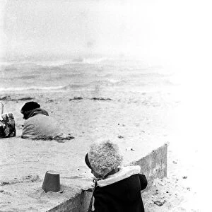
Blood clot on plaster, SEM
Blood clot on plaster. Coloured scanning electron micrograph (SEM) of blood clotting on the surface of a sticking plaster used to dress a small cut. Red blood cells (erythrocytes) are trapped in filaments of fibrin protein (brown). The fibres of the absorbent material of the plaster are also seen (grey). When an injury occurs, platelets (not seen) in the blood stimulate the formation of fibrin filaments, which enmesh platelets and red and white blood cells. The fibrin contracts around them to form a solid clot. Sticking plasters are used to help stop bleeding, and to keep an injury clean and dry during healing. Magnification: x830 when printed 10cm wide
Science Photo Library features Science and Medical images including photos and illustrations
Media ID 6421178
© STEVE GSCHMEISSNER/SCIENCE PHOTO LIBRARY
Blood Blood Clot Circulatory Clot Clotting Coagulated Coagulation Coloured Sem Dressing Erythrocyte Erythrocytes Fibrin Haematology Mesh Physiology Plaster Process Sticking Sticking Plaster Strand Strands Treatment Vascular Circulation Red Cells
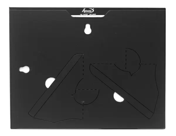
7"x5" Glass Mount
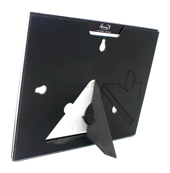
Wall mounted or free-standing, these black edged glass frames feature a smooth chamfered edge and a stylish black border (on back face of the glass). Manufactured from 4mm thick glass, Glass Mounts are a durable, professional way of displaying and protecting your prints. Your 7x5 print is slotted into the back of the frame so can easily be changed if needed.
Tempered Glass Mounts are ideal for wall display, plus the smaller sizes can also be used free-standing via an integral stand
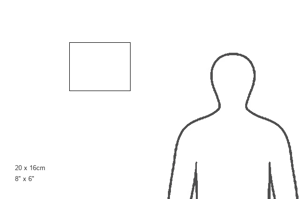
Estimated Image Size (if not cropped) is 17.7cm x 12.7cm (7" x 5")
Estimated Product Size is 20.3cm x 16.2cm (8" x 6.4")
These are individually made so all sizes are approximate
Artwork printed orientated as per the preview above, with landscape (horizontal) orientation to match the source image.
FEATURES IN THESE COLLECTIONS
EDITORS COMMENTS
This print showcases the intricate process of blood clotting on a sticking plaster used to dress a small cut. The vibrant colors in this coloured scanning electron micrograph (SEM) highlight the fascinating details of this physiological phenomenon. Red blood cells, depicted as erythrocytes, are entangled within filaments of fibrin protein, giving them a striking brown hue. Additionally, the absorbent material of the plaster is visible in shades of grey. When an injury occurs, platelets play a crucial role by stimulating the formation of fibrin filaments that ensnare platelets and red and white blood cells alike. As these filaments contract around them, they solidify into a cohesive clot. Sticking plasters serve two essential purposes: halting bleeding and maintaining cleanliness during the healing process. At an impressive magnification level of x830 when printed 10cm wide, this image provides viewers with an up-close look at one aspect of our body's remarkable ability to heal itself. It offers insights into both human anatomy and physiology while shedding light on various aspects such as circulation and coagulation. Science Photo Library has captured this extraordinary moment in haematology using scanning electron microscopy techniques. This photograph serves as a reminder not only of our body's incredible resilience but also highlights the importance of medical advancements that aid in wound care and recovery processes.
MADE IN THE UK
Safe Shipping with 30 Day Money Back Guarantee
FREE PERSONALISATION*
We are proud to offer a range of customisation features including Personalised Captions, Color Filters and Picture Zoom Tools
SECURE PAYMENTS
We happily accept a wide range of payment options so you can pay for the things you need in the way that is most convenient for you
* Options may vary by product and licensing agreement. Zoomed Pictures can be adjusted in the Basket.