Fraud Blocker Skip to main content

Clot Collection

"Unraveling the Intricacies of Clot

Background imageClot Collection: Blood coagulation cascade, artwork C016 / 9873

Blood coagulation cascade, artwork C016 / 9873
Blood coagulation cascade. Artwork of the biochemical cascade of blood chemicals and proteins during blood clotting (coagulation). The blood vessel and its layered wall is at upper left

Background imageClot Collection: Liver cirrhosis

Liver cirrhosis. Artwork taken from Sir Robert Carswells textbook Pathological Anatomy, published 1838. Cirrhosis is a disease in which the liver tissue is replaced with scar tissue

Background imageClot Collection: Blood clot on plaster, SEM

Blood clot on plaster, SEM
Blood clot on plaster. Coloured scanning electron micrograph (SEM) of blood clotting on the surface of a sticking plaster used to dress a small cut

Background imageClot Collection: Fans in the Loan Exhibition in the South Kensington Museum (engraving)

Fans in the Loan Exhibition in the South Kensington Museum (engraving)
1104451 Fans in the Loan Exhibition in the South Kensington Museum (engraving) by English School, (19th century); Private Collection; (add.info)

Background imageClot Collection: Fireworks Safety Campaign ahead of Bonfire Night, 1st November 1968

Fireworks Safety Campaign ahead of Bonfire Night, 1st November 1968. Lancashire Home Safety Committees

Background imageClot Collection: Stent inside of an artery, illustration

Stent inside of an artery, illustration

Background imageClot Collection: Banks of the River (Les Bords de riviere), 1897. Creators: Alfred Sisley

Banks of the River (Les Bords de riviere), 1897. Creators: Alfred Sisley
Banks of the River (Les Bords de riviere), 1897

Background imageClot Collection: Woman Carrying a Birthday Cake

Woman Carrying a Birthday Cake
http://csaimages.com/images/istockprofile/csa_vector_dsp.jpg

Background imageClot Collection: Pulmonary embolism, pathway of embolus to the heart and lungs

Pulmonary embolism, pathway of embolus to the heart and lungs

Background imageClot Collection: Ischaemia, digital angiogram

Ischaemia, digital angiogram
image d angiographie numerisee( non soustraite) de la main gauche, en vue de face, par catheterisme hyperselectif des arteres du membre superieur gauche, sous anasthesie generale

Background imageClot Collection: Blocked artery, computer artwork

Blocked artery, computer artwork
Blocked artery. Computer artwork of a clot of blood cells (thrombus, red) in an artery affected by atherosclerosis. This is a narrowing of an artery due to fatty deposits (yellow) on its inner walls

Background imageClot Collection: Antoine Bart. Clot

Antoine Bart. Clot
ANTOINE BARTHELEMY CLOT known as CLOT-BEY French medical in Egypt in later life. Date: 1793 - 1868

Background imageClot Collection: Brain stoke, artwork F008 / 0227

Brain stoke, artwork F008 / 0227
Brain stoke, computer artwork

Background imageClot Collection: Coloured SEM of an early blood clot forming

Coloured SEM of an early blood clot forming
Blood clot. Coloured Scanning Electron Micrograph of an early blood clot. The red blood cells are forming rouleaux (rolls) as they become enmeshed in a tangle of fibrin fibres (yellow)

Background imageClot Collection: Blood clot, light micrograph

Blood clot, light micrograph
Blood clot. Light micrograph of a section through an arterial thrombus. Blood clots can form in an intact vessel that contains fatty deposits or one that is inflamed

Background imageClot Collection: Thrombin protein, molecular model F006 / 9603

Thrombin protein, molecular model F006 / 9603
Thrombin protein, molecular model. Thrombin is an enzyme involved in the blood coagulation (clotting) process. It converts fibrinogen (a soluble plasma glycoprotein synthesised in the liver)

Background imageClot Collection: Coronary stenosis after treatment, X-ray

Coronary stenosis after treatment, X-ray
Coronary stenosis after treatment. Angiogram of the left coronary artery in the heart of a 60-year-old patient after balloon catheterisation

Background imageClot Collection: Coronary stenosis treatment, X-rays

Coronary stenosis treatment, X-rays
Coronary stenosis treatment. Coloured angiograms showing the left coronary artery in the heart of a 60-year-old patient before (top left)

Background imageClot Collection: Brain haemorrhage, CT scans C018 / 0569

Brain haemorrhage, CT scans C018 / 0569
Brain haemorrhage. Coloured computed tomography (CT) scans of the brain of a 49 year old patient with a subdural haematoma (blue)

Background imageClot Collection: Thrombin protein, molecular model C015 / 7074

Thrombin protein, molecular model C015 / 7074
Thrombin protein, molecular model. Thrombin is an enzyme involved in the blood coagulation (clotting) process. It converts fibrinogen (a soluble plasma glycoprotein synthesised in the liver)

Background imageClot Collection: Pulmonary embolism, X-ray C017 / 7802

Pulmonary embolism, X-ray C017 / 7802
Pulmonary embolism. X-ray of the chest of a 72 year old male patient with a pulmonary embolism. This is a blockage of the pulmonary artery

Background imageClot Collection: Coloured SEM of a blood clot in coronary artery

Coloured SEM of a blood clot in coronary artery
Thrombus. Coloured Scanning Electron Micrograph (SEM) of a thrombus (blood clot) inside a coronary artery of the human heart

Background imageClot Collection: Brain haemorrhage, CT scan

Brain haemorrhage, CT scan

Background imageClot Collection: Coloured SEM of a blood clot due to an injury

Coloured SEM of a blood clot due to an injury

Background imageClot Collection: Computer artwork of a blood clot

Computer artwork of a blood clot
Blood clot. Computer artwork of a blood clot, showing disc-like red blood cells and fibrin. Fibrin (yellow fibres) is a long insoluble protein produced from the blood protein fibrinogen

Background imageClot Collection: Coloured SEM of red blood cells forming a clot

Coloured SEM of red blood cells forming a clot
Blood clot. Coloured Scanning Electron Micrograph (SEM) showing the formation of a blood clot. Red blood cells (erythrocytes)

Background imageClot Collection: Illustration of a dissolving blood clot (thrombus)

Illustration of a dissolving blood clot (thrombus)
Dissolving blood clot. Illustration of a human blood clot (thrombus) in the process of dissolving. At upper centre is the blood clot

Background imageClot Collection: Healing wound, SEM

Healing wound, SEM
Healing skin wound, coloured scanning electron micrograph (SEM). A blood vessel is leaking at left. At right, red blood cells (or erythrocytes)

Background imageClot Collection: Deep vein thrombosis, artwork

Deep vein thrombosis, artwork
Deep vein thrombosis. Computer artwork showing blood clots in a leg (brown lumps, bottom right), caused by deep vein thrombosis (DVT), moving to blood vessels in the lungs (blue, top right)

Background imageClot Collection: Coloured angiogram of embolus blocking arm artery

Coloured angiogram of embolus blocking arm artery

Background imageClot Collection: Illustration of blood cells & fibrin in a thrombus

Illustration of blood cells & fibrin in a thrombus
Thrombus. Illustration of a thrombus, an abnormal blood clot formed inside a vein or artery. Red blood cells appear trapped inside a network of fibres of the insoluble protein fibrin

Background imageClot Collection: SEM of blood clot

SEM of blood clot
Thrombosis. False-colour scanning electron micrograph (SEM) of a thrombus (clot) of red blood cells inside a blood vessel

Background imageClot Collection: Blocked coronary artery, artwork

Blocked coronary artery, artwork
Blocked coronary artery. Computer artwork of a clot (red) in a coronary artery. The clot has been caused by atherosclerosis, the build up of fatty plaques (yellow) on the artery wall

Background imageClot Collection: Coronary thrombosis, light micrograph

Coronary thrombosis, light micrograph
Coronary thrombosis. Light micrograph of a section through a thrombus (blood clot) in the coronary artery of the heart. The clot is packed full of red blood cells (red)

Background imageClot Collection: Blood clot, artwork

Blood clot, artwork

Background imageClot Collection: Subarachnoid haemorrhage, MRI scan

Subarachnoid haemorrhage, MRI scan
Subarachnoid haemorrhage. Magnetic resonance imaging (MRI) scan of an axial section through the brain of a patient with a subarachnoid haematoma (light grey, centre left)

Background imageClot Collection: Activated platelets, SEM

Activated platelets, SEM
Activated platelets. Coloured scanning electron micrograph (SEM) of activated platelets, or thrombocytes, starting to adhere to exposed sub-endothelium on the inner surface of a vein

Background imageClot Collection: Blood clotting, SEM

Blood clotting, SEM
Blood clotting. Coloured scanning electron micrograph (SEM) of human red blood cells (erythrocytes, red) and platelets (thrombocyte, white) forming a blood clot

Background imageClot Collection: Red blood cells and platelets, SEM

Red blood cells and platelets, SEM
Red blood cells and platelets. Coloured scanning electon micrograph (SEM) of human erythrocytes (red blood cells) and a platelet aggregate (purple)

Background imageClot Collection: Activated platelets, artwork

Activated platelets, artwork
Activated platelets. Artwork of platelets (thrombocytes) that have been activated. Platelets are part of the blood. When a blood vessel is damaged

Background imageClot Collection: Thrombin protein

Thrombin protein

Background imageClot Collection: Thrombin protein, secondary structure

Thrombin protein, secondary structure
Thrombin protein, computer model. Thrombin is a protein involved in the blood coagulation (clotting) process. It converts fibrinogen (a soluble plasma glycoprotein synthesised in the liver)

Background imageClot Collection: NovoSeven clotting protein molecule

NovoSeven clotting protein molecule
NovoSeven clotting protein, molecular model. NovoSeven is the brand name for an artificial version of the natural blood protein Factor VII, or proconvertin

Background imageClot Collection: Fibrin strands, artwork

Fibrin strands, artwork
Fibrin strands. Computer artwork depicting a number of fibrin strands with their ends embedded in the membrane of a cell. Fibrin is composed of fibrinogen proteins

Background imageClot Collection: Silent Film Still: Beach

Silent Film Still: Beach

Background imageClot Collection: Curiosities and ingenuities from the Patent Office; Fly clot

Curiosities and ingenuities from the Patent Office; Fly clot
Illustration showing protective clothing for horses and other animals to prevent them being bothered by flies and other insects


All Professionally Made to Order for Quick Shipping

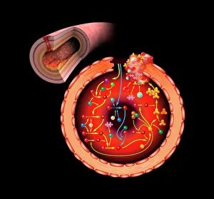
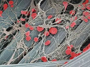
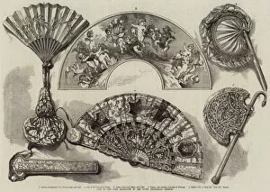
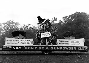
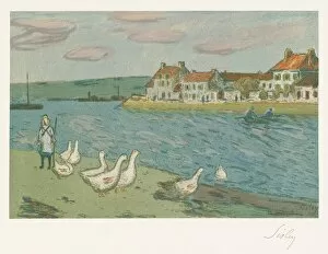
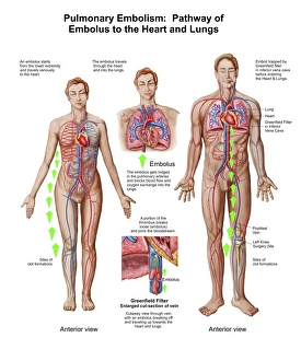
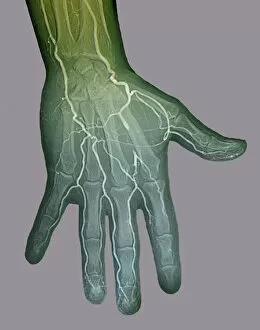
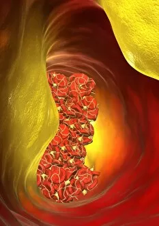
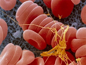
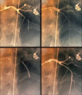
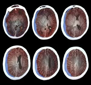
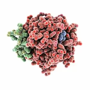
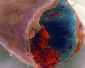
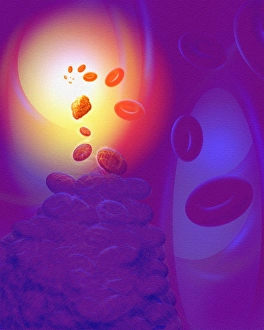
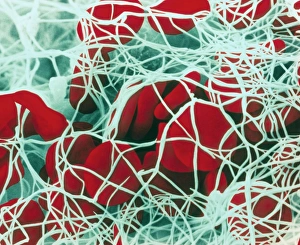
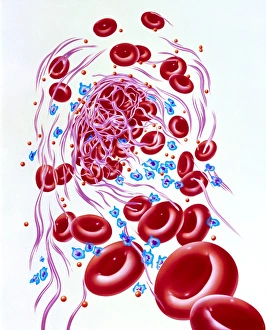
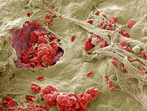
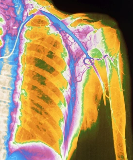
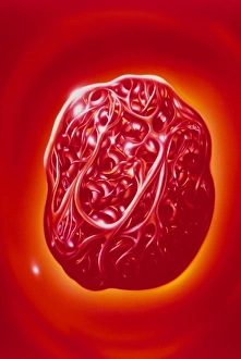
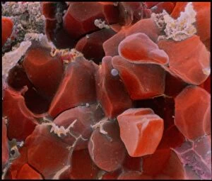
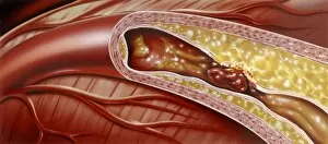
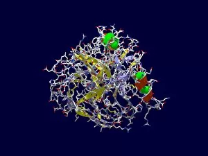
"Unraveling the Intricacies of Clot: From Blood Coagulation Cascade to Liver Cirrhosis" Discover the fascinating world of clotting with artwork C016/9873 depicting the intricate blood coagulation cascade. Dive into the complexities of liver cirrhosis and its impact on clot formation. Witness a mesmerizing SEM image capturing a blood clot on plaster, showcasing the microscopic beauty within our bodies. Step back in time as you join fans at the Loan Exhibition in South Kensington Museum through an enchanting engraving, reminding us that art can be found even in unexpected places like clots. Explore medical advancements with an illustration displaying a stent inside an artery, revolutionizing treatments for cardiovascular conditions. Immerse yourself in Alfred Sisley's masterpiece "Banks of the River (Les Bords de riviere), 1897, " where nature's tranquility contrasts with life's turbulent clots. Celebrate joyous moments like birthdays while admiring Antoine Bart Clot's artistic prowess captured in "Woman Carrying a Birthday Cake. " Delve into history as we remember Antoine Bart Clot, who left his mark on art and culture. Promote safety awareness during Bonfire Night by recalling the Fireworks Safety Campaign from November 1st, 1968 – let's prevent accidents and ensure celebrations remain free from harm. Educate yourself about pulmonary embolism by tracing its pathway to the heart and lungs, understanding how dangerous they can become life-threatening if not addressed promptly. Witness ischaemia through a digital angiogram, shedding light on vascular blockages caused by clots and their consequences on tissue health.