Fraud Blocker Skip to main content

Clotting Collection

"Unraveling the Intricate Dance of Clotting

Background imageClotting Collection: Blood clot, SEM

Blood clot, SEM
Blood clot, coloured scanning electron micrograph (SEM). Red blood cells are red and fibrin protein strands are green. Platelets are at bottom right

Background imageClotting Collection: Blood coagulation cascade, artwork C016 / 9873

Blood coagulation cascade, artwork C016 / 9873
Blood coagulation cascade. Artwork of the biochemical cascade of blood chemicals and proteins during blood clotting (coagulation). The blood vessel and its layered wall is at upper left

Background imageClotting Collection: Liver cirrhosis

Liver cirrhosis. Artwork taken from Sir Robert Carswells textbook Pathological Anatomy, published 1838. Cirrhosis is a disease in which the liver tissue is replaced with scar tissue

Background imageClotting Collection: Blood clot on plaster, SEM

Blood clot on plaster, SEM
Blood clot on plaster. Coloured scanning electron micrograph (SEM) of blood clotting on the surface of a sticking plaster used to dress a small cut

Background imageClotting Collection: Ischaemia, digital angiogram

Ischaemia, digital angiogram
image d angiographie numerisee( non soustraite) de la main gauche, en vue de face, par catheterisme hyperselectif des arteres du membre superieur gauche, sous anasthesie generale

Background imageClotting Collection: Blood clot, artwork C016 / 4619

Blood clot, artwork C016 / 4619
Blood clot in a blood vessel, computer artwork. Red blood cells (erythrocytes) are trapped within a fibrin protein mesh (cream)

Background imageClotting Collection: White blood cells and platelets, SEM C016 / 3099

White blood cells and platelets, SEM C016 / 3099
White blood cells and platelets. Coloured scanning electron micrograph (SEM) of platelets (green) with a variety of different white blood cells (leucocytes, yellow)

Background imageClotting Collection: White blood cells and platelets, SEM C016 / 3098

White blood cells and platelets, SEM C016 / 3098
White blood cells and platelets. Coloured scanning electron micrograph (SEM) of platelets (purple) with a variety of different white blood cells (leucocytes, blue)

Background imageClotting Collection: Coagulation factor complex molecule C014 / 0139

Coagulation factor complex molecule C014 / 0139
Coagulation factor complex molecule. Molecular model showing the interaction between coagulation factor VIII (FVIII, pink, blue and yellow) and factor IXa (FIXa, cream and grey)

Background imageClotting Collection: Thrombosed blood vessel, artwork C013 / 4649

Thrombosed blood vessel, artwork C013 / 4649
Thrombosed blood vessel. Computer artwork of a blood clot (thrombus) formed on the internal wall of a blood vessel

Background imageClotting Collection: Blood platelets

Blood platelets. Coloured scanning electron micro- graph (SEM) of activated blood platelets. Platelet cells (thrombocytes) are formed in the bone marrow

Background imageClotting Collection: Blood clot, SEM P260 / 0123

Blood clot, SEM P260 / 0123
Blood clot. Coloured scanning electron micrograph (SEM) of a blood clot. The red blood cells (erythrocytes) are trapped in filaments of fibrin protein (pink)

Background imageClotting Collection: Integrin and fibrinogen complex molecule F006 / 9649

Integrin and fibrinogen complex molecule F006 / 9649
Integrin and fibrinogen complex. Molecular model of integrin alpha-II beta-3 complexed with fibrinogen. Integrin alpha-II beta-3 is a transmembrane protein found on platelets

Background imageClotting Collection: Blood clotting factor VIII molecule C014 / 0406

Blood clotting factor VIII molecule C014 / 0406
Blood clotting factor VIII, molecular model showing the secondary structure. Factor VIII, also known as anti-haemophilic factor, is an essential blood clotting protein in humans

Background imageClotting Collection: Thrombin protein, molecular model F006 / 9603

Thrombin protein, molecular model F006 / 9603
Thrombin protein, molecular model. Thrombin is an enzyme involved in the blood coagulation (clotting) process. It converts fibrinogen (a soluble plasma glycoprotein synthesised in the liver)

Background imageClotting Collection: Thrombin complexed with fibrinogen F006 / 9561

Thrombin complexed with fibrinogen F006 / 9561
Thrombin complexed with fibrinogen, molecular model. The thrombin molecules (beige, right and left) are bound to the central part of the fibrinogen molecule (down centre)

Background imageClotting Collection: Blood clot, SEM C016 / 9088

Blood clot, SEM C016 / 9088
Blood clot. Coloured scanning electron micrograph (SEM) of a section through a blood clot (thrombus) in an arteriole (small blood vessel), showing red blood cells (red) in a fibrin mesh (green)

Background imageClotting Collection: Blood clot, SEM C016 / 9089

Blood clot, SEM C016 / 9089
Blood clot. Coloured scanning electron micrograph (SEM) of a section through a blood clot (thrombus) in an arteriole (small blood vessel), showing red blood cells (red) in a fibrin mesh (bright pink)

Background imageClotting Collection: Coronary blood clot, SEM C016 / 9133

Coronary blood clot, SEM C016 / 9133
Coronary blood clot. Coloured scanning electron micrograph (SEM) of a blood clot (thrombus) in the coronary artery, showing red blood cells (purple) in a fibrin mesh (threads)

Background imageClotting Collection: Thrombin complexed with fibrinogen C015 / 7148

Thrombin complexed with fibrinogen C015 / 7148
Thrombin complexed with fibrinogen, molecular model. The thrombin molecules (left and right, purple and green) are bound to the central part of the fibrinogen molecule (centre, multiple colours)

Background imageClotting Collection: Thrombin complexed with fibrinogen C015 / 7149

Thrombin complexed with fibrinogen C015 / 7149
Thrombin complexed with fibrinogen, molecular model. The thrombin molecules (left and right, brown and pink) are bound to the central part of the fibrinogen molecule (centre, multiple colours)

Background imageClotting Collection: Blood clotting factor VIII molecule C014 / 0407

Blood clotting factor VIII molecule C014 / 0407
Blood clotting factor VIII, molecular model showing the secondary structure. Factor VIII, also known as anti-haemophilic factor, is an essential blood clotting protein in humans

Background imageClotting Collection: Coagulation factor complex molecule C014 / 0409

Coagulation factor complex molecule C014 / 0409
Coagulation factor complex molecule. Molecular model showing the interaction between coagulation factor VIII (FVIII, pink, blue and yellow), factor IXa (FIXa)

Background imageClotting Collection: Blood clotting factor VIII molecule C014 / 0408

Blood clotting factor VIII molecule C014 / 0408
Blood clotting factor VIII, molecular model. Factor VIII, also known as anti-haemophilic factor, is an essential blood clotting protein in humans

Background imageClotting Collection: Coagulation factor complex molecule C014 / 0410

Coagulation factor complex molecule C014 / 0410
Coagulation factor complex molecule. Molecular model showing the interaction between coagulation factor VIII (FVIII, pink, blue and yellow), factor IXa (FIXa)

Background imageClotting Collection: Blood clot, SEM C014 / 0381

Blood clot, SEM C014 / 0381
Blood clot. Coloured scanning electron micrograph (SEM) of a blood clot (thrombus) in an arteriole (small blood vessel) of a salivary gland

Background imageClotting Collection: Blood clot, SEM C014 / 0380

Blood clot, SEM C014 / 0380
Blood clot. Coloured scanning electron micrograph (SEM) of a blood clot (thrombus) in an arteriole (small blood vessel) of a salivary gland

Background imageClotting Collection: Thrombin protein, molecular model C015 / 7074

Thrombin protein, molecular model C015 / 7074
Thrombin protein, molecular model. Thrombin is an enzyme involved in the blood coagulation (clotting) process. It converts fibrinogen (a soluble plasma glycoprotein synthesised in the liver)

Background imageClotting Collection: Blood clot, artwork C016 / 4618

Blood clot, artwork C016 / 4618
Blood clot, computer artwork. Red blood cells (erythrocytes) are trapped within a fibrin protein mesh (cream). The fibrin mesh is formed in response to chemicals secreted by platelets (white)

Background imageClotting Collection: Blood clot, artwork C016 / 4620

Blood clot, artwork C016 / 4620
Fortification agate. Section through a fortification agate from Scurdie Ness, Scotland, UK. Agate is an aggregate of different forms of silica (SiO2) within a geode

Background imageClotting Collection: White blood cells, SEM C016 / 3102

White blood cells, SEM C016 / 3102
White blood cells. Coloured scanning electron micrograph (SEM) of white blood cells (leucocytes) from a sample of peripheral blood

Background imageClotting Collection: Blood cells, SEM C016 / 3101

Blood cells, SEM C016 / 3101
Blood cells. Coloured scanning electron micrograph (SEM) of a variety of different white blood cells (leucocytes, blue/purple) with two red blood cells (erythrocytes)

Background imageClotting Collection: Blood cells, SEM C016 / 3100

Blood cells, SEM C016 / 3100
Blood cells. Coloured scanning electron micrograph (SEM) of a variety of different white blood cells (leucocytes, yellow/green) with two red blood cells (erythrocytes)

Background imageClotting Collection: Macrophage and platelets, SEM C016 / 3094

Macrophage and platelets, SEM C016 / 3094
Macrophage and platelets. Coloured scanning electron micrograph (SEM) of platelets (green) attached to a macrophage white blood cell (turquoise)

Background imageClotting Collection: Platelets, SEM C016 / 3091

Platelets, SEM C016 / 3091
Platelets, coloured scanning electron micrograph (SEM). Platelets, or thrombocytes, are fragments of white blood cells. When blood clots

Background imageClotting Collection: Blood clot C013 / 7129

Blood clot C013 / 7129
Blood clot. Coloured scanning electron micrograph (SEM) of a blood clot (centre) forming in connective tissue. Red blood cells (erythrocytes)

Background imageClotting Collection: Blood clot C013 / 7128

Blood clot C013 / 7128
Blood clot. Coloured scanning electron micrograph (SEM) of a blood clot (centre) forming in connective tissue. Red blood cells (erythrocytes)

Background imageClotting Collection: Blood cells, artwork C013 / 4726

Blood cells, artwork C013 / 4726
Blood cells. Computer artwork of red blood cells (erythrocytes, red), white blood cells (leucocytes, white), and platelets (thrombocytes, yellow)

Background imageClotting Collection: Blood cells, artwork C013 / 4707

Blood cells, artwork C013 / 4707
Blood cells. Computer artwork of red blood cells (erythrocytes, red), white blood cells (leucocytes, white), and platelets (thrombocytes, yellow)

Background imageClotting Collection: F / col TEM Unactivated blood platelets

F / col TEM Unactivated blood platelets
False-colour transmission electron micrograph of unactivated blood platelets & some red blood cells (bottom left). Platelets (thrombocytes) are small

Background imageClotting Collection: Fibrin mesh in a blood clot

Fibrin mesh in a blood clot. Coloured scanning electron micrograph of fibrin fibres in a blood clot. When blood vessels become damaged

Background imageClotting Collection: Blood clots, SEM

Blood clots, SEM
Blood clots. Coloured scanning electron micrograph (SEM) of small blood clots (red) in a blood vessel. The clots consist of red blood cells, white blood cells, and platelets

Background imageClotting Collection: Platelets in a blood clot

Platelets in a blood clot. Coloured scanning electron micrograph (SEM) of platelet cells (thrombocytes) in a blood clot. They are oval shaped

Background imageClotting Collection: F / col TEM of section through blood platelets

F / col TEM of section through blood platelets
False colour transmission electron micrograph of a section through activated blood platelets. Platelets (thrombocytes) are formed in the bone marrow, and circulate in blood in large numbers

Background imageClotting Collection: Blood clot crystals, SEM

Blood clot crystals, SEM
Blood clot crystals. Coloured scanning electron micrograph (SEM) of crystals of albumin from a blood clot. Albumin is the most abundant protein in the blood plasma

Background imageClotting Collection: Coloured SEM of red blood cells forming a clot

Coloured SEM of red blood cells forming a clot
Blood clot. Coloured Scanning Electron Micrograph (SEM) showing the formation of a blood clot. Red blood cells (erythrocytes)

Background imageClotting Collection: Illustration of a dissolving blood clot (thrombus)

Illustration of a dissolving blood clot (thrombus)
Dissolving blood clot. Illustration of a human blood clot (thrombus) in the process of dissolving. At upper centre is the blood clot

Background imageClotting Collection: Blood clot

Blood clot
False-colour scanning electron micrograph (SEM) of a blood clot. Red blood cells have been trapped by a web of thin yellow-white strands of fibrin


All Professionally Made to Order for Quick Shipping

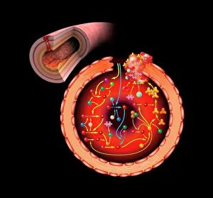
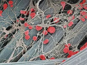
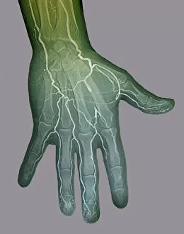
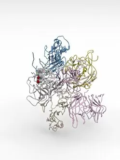
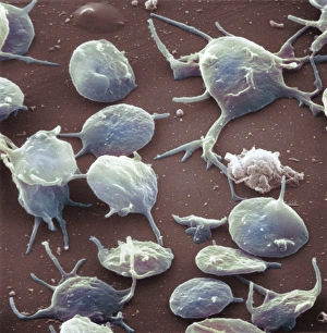
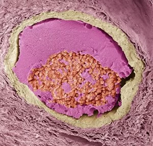
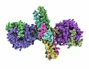
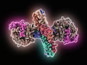
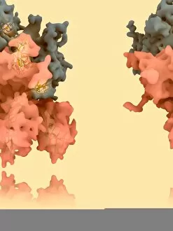
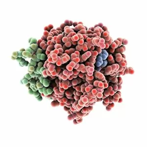
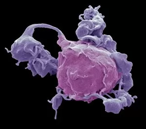
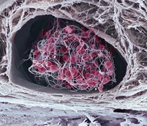
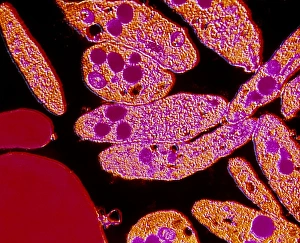
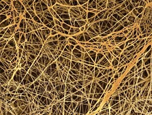
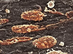
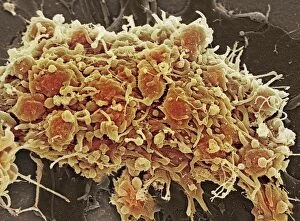
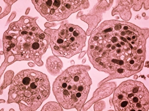
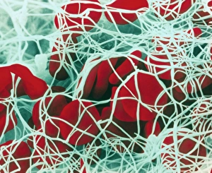
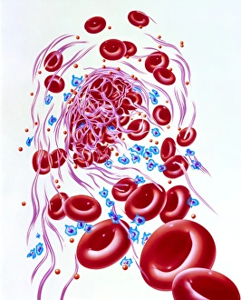
"Unraveling the Intricate Dance of Clotting: Exploring the Blood Coagulation Cascade" Intriguing artwork C016 / 9873 takes us on a visual journey into the complex process of blood clotting. Liver cirrhosis, a condition that affects liver function, can disrupt this delicate balance and lead to abnormal clot formation. SEM images reveal the intricate structure of a blood clot on plaster (Blood clot on plaster, SEM) and offer a closer look at individual platelets coming together to form a solid mass (Platelets, SEM). Ischaemia is captured in stunning detail through digital angiogram imagery (Ischaemia, digital angiogram), showcasing how inadequate blood flow can result in tissue damage due to insufficient oxygen supply. Artwork C016 / 4619 beautifully illustrates the formation of a blood clot as it obstructs normal circulation within our bodies. SEM images C016 / 3099 and C016 / 3098 provide captivating glimpses into the collaboration between white blood cells and platelets during coagulation - an essential defense mechanism against excessive bleeding. The coagulation factor complex molecule (Coagulation factor complex molecule C014 / 0139) plays a crucial role in orchestrating this intricate dance by activating various proteins involved in clot formation. Thrombosed blood vessels are brought to life through artwork C013 / 4649, emphasizing their potential consequences if left untreated or unmanaged. Finally, SEM image P260 / 0123 showcases another mesmerizing close-up view of a formed blood clot – highlighting its unique composition and structure. Through these captivating visuals and scientific insights, we gain deeper appreciation for the remarkable phenomenon known as "clotting, " which safeguards our bodies from excessive bleeding while reminding us of its critical importance in maintaining overall health.