
Framed Print > Arts > Artists > J > Jacob Jacobs
Framed Print : Cranial nerves


![]()
Framed Photos from Science Photo Library
Cranial nerves
Cranial nerves, historical anatomical artwork. This neck and upper torso have been dissected to show the paths of the twelve cranial nerves (white). The cranial nerves emerge in the brainstem at the base of the brain, and mediate the movements and senses of the face and head. The vagus nerve, the 10th cranial nerve (central, white), extends to the abdomen, innervating the larynx, heart (lower, orange) and digestive system. This illustration is taken from the nineteenth-century French textbook, the Atlas of Human Anatomy and Surgery, by J. M. Bourgery and N. H. Jacob
Science Photo Library features Science and Medical images including photos and illustrations
Media ID 6448329
© MEHAU KULYK/SCIENCE PHOTO LIBRARY
Aorta Atlas Of Human Anatomy Brain Stem Bronchi Bronchus Central Nervous System Cranial Nerve Dissected Dissection French Innervate Innervating J M Bourgery Lung Lungs N H Jacob Nerve Nerves Peripheral Nervous System Rhombencephalon Spinal Cord Surgery Surgical Thoracic Thorax Vagus Nerve Dissect Neurological Neurology Section
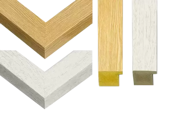
14"x12" (38x32cm) Modern Frame
Discover the intricacies of human anatomy with our Media Storehouse Framed Prints featuring the captivating image of "Cranial Nerves" by Science Photo Library. This historical anatomical artwork offers a fascinating glimpse into the complex network of the twelve cranial nerves, delicately illustrated against the backdrop of a meticulously dissected neck and upper torso. Each print is expertly framed to preserve the intricate details and vibrant colors of the original image, making it a stunning addition to any home or office space. Ignite your curiosity and deepen your understanding of the intricacies of the human body with our Framed Prints collection.
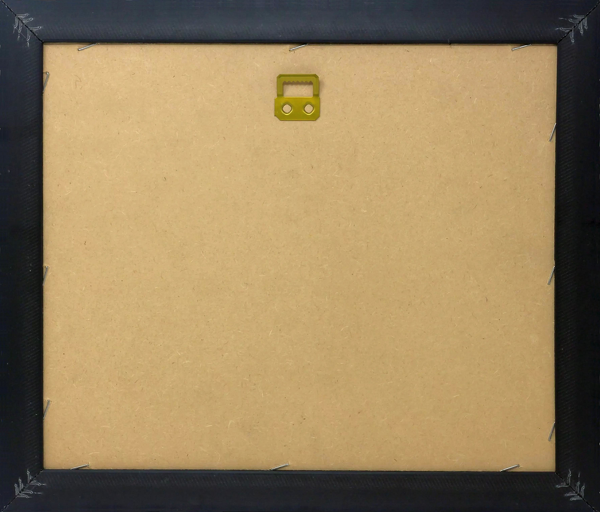
Wood effect frame, card mounted, 10x8 archival quality photo print. Overall outside dimensions 14x12 inches (38x32cm). Environmentally and ozone friendly, 40mm wide x 15mm Polycore® moulding has the look of real wood, is durable and light and easy to hang. Biodegradable and made with non-chlorinated gases (no toxic fumes) it is efficient; producing 100 tons of polystyrene can save 300 tons of trees! Prints are glazed with lightweight, shatterproof, optical clarity acrylic (providing the same general protection from the environment as glass). The back is stapled hardboard with a sawtooth hanger attached. Note: To minimise original artwork cropping, for optimum layout, and to ensure print is secure, the visible print may be marginally smaller
Contemporary Framed and Mounted Prints - Professionally Made and Ready to Hang
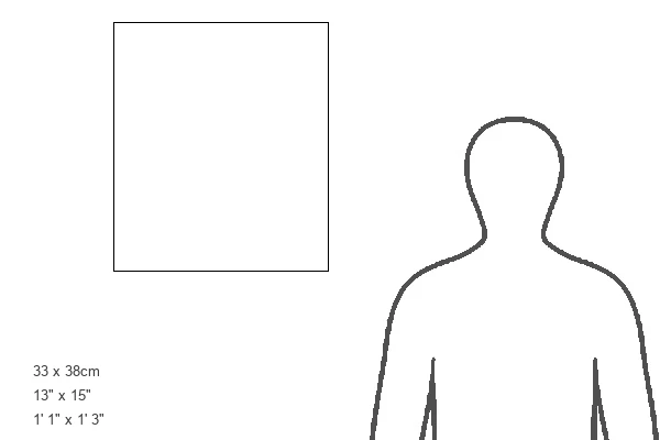
Estimated Image Size (if not cropped) is 20.2cm x 24.4cm (8" x 9.6")
Estimated Product Size is 32.5cm x 37.6cm (12.8" x 14.8")
These are individually made so all sizes are approximate
Artwork printed orientated as per the preview above, with portrait (vertical) orientation to match the source image.
EDITORS COMMENTS
This print showcases a historical anatomical artwork depicting the intricate network of cranial nerves within the human body. The image reveals a dissected neck and upper torso, meticulously illustrating the paths of the twelve cranial nerves in white. These vital nerves emerge from the brainstem at the base of the brain, playing a crucial role in mediating movements and senses related to the face and head. One particular nerve that stands out is the vagus nerve, also known as the 10th cranial nerve. This central white structure extends beyond its counterparts, reaching all the way down to innervate various organs such as the larynx, heart (depicted in lower orange), and digestive system located in our abdomen. Its extensive reach demonstrates its significance in regulating essential bodily functions. The artwork originates from an esteemed nineteenth-century French textbook called "Atlas of Human Anatomy and Surgery" authored by J. M. Bourgery and N. H. Jacob. This masterpiece not only serves as a testament to their expertise but also provides us with valuable insights into early surgical practices. With its detailed portrayal of cut-out sections showcasing organs like lungs, bronchi, spinal cord, thorax, and even highlighting structures like aorta and rhombencephalon (hindbrain), this illustration offers an invaluable glimpse into our complex biological makeup. As we delve into this mesmerizing piece of history captured by Science Photo Library's collection, we are reminded once again of how far we have come in understanding our own bodies through advancements in surgery, biology, neurology - ultimately unraveling countless mysteries hidden within our central nervous system.
MADE IN THE UK
Safe Shipping with 30 Day Money Back Guarantee
FREE PERSONALISATION*
We are proud to offer a range of customisation features including Personalised Captions, Color Filters and Picture Zoom Tools
SECURE PAYMENTS
We happily accept a wide range of payment options so you can pay for the things you need in the way that is most convenient for you
* Options may vary by product and licensing agreement. Zoomed Pictures can be adjusted in the Basket.

