
Atlas Of Human Anatomy Collection
The "Atlas of Human Anatomy" is a comprehensive guide that delves into the intricacies of the human body


All Professionally Made to Order for Quick Shipping
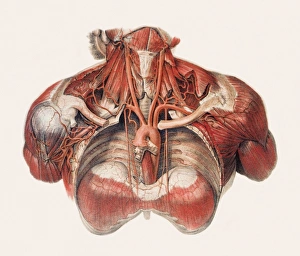
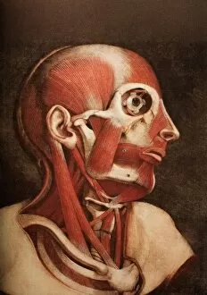
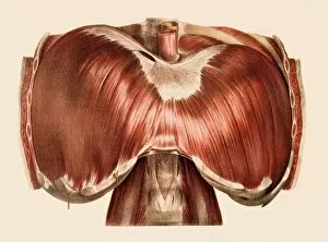
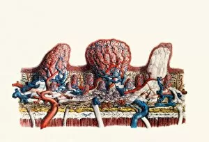
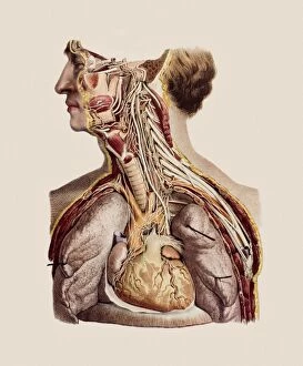
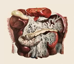
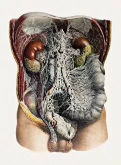
The "Atlas of Human Anatomy" is a comprehensive guide that delves into the intricacies of the human body, providing an in-depth exploration of various systems and structures. From the protective layers surrounding our brain known as meninges to the complex network of blood vessels found in our chest and neck, this atlas leaves no stone unturned. One section focuses on the muscles that make up our head and neck, shedding light on their functions and interactions. Another highlights the diaphragm, a vital muscle responsible for respiration. The papillae on our tongue are also examined closely, revealing fascinating details about taste perception. For those interested in neurological anatomy, this atlas offers detailed insights into cranial nerves and brain structure. It provides a roadmap to understanding how these intricate networks facilitate communication within our bodies. Furthermore, it explores surgical procedures related to bowel surgery, offering valuable information for medical professionals involved in such interventions. Additionally, it covers abdominal organs extensively – from their composition to their interplay with kidneys, nerves, and blood vessels. Whether you're a medical student seeking knowledge or simply curious about what lies beneath your skin's surface, this "Atlas of Human Anatomy" serves as an indispensable resource. Its meticulous illustrations and informative descriptions provide a captivating journey through the complexities of our remarkable bodies.