Framed Print > Sport > Cycling
Framed Print : Cell division, fluorescent micrograph


![]()
Framed Photos from Science Photo Library
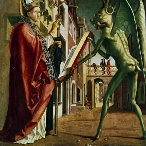
Cell division, fluorescent micrograph
Cell division. Immunofluorescent light micrograph of a human epithelial cell (centre) during the interphase stage of mitosis. Mitosis is the cycle of replication and division by which new body cells are formed. Here, the chromosomes of the parent cell (blue) have begun to duplicate and pair up, and the microtubules (green) have formed. The microtubules will pull the chromosome pairs apart to opposite poles of the cell, in a process called cytokinesis. When the cell has split, two identical daughter cells are formed
Science Photo Library features Science and Medical images including photos and illustrations
Media ID 6326105
© DR TORSTEN WITTMANN/SCIENCE PHOTO LIBRARY
Cell Biology Cell Division Cellular Chromosome Chromosomes Cycle Cytological Cytology Cytoplasm Daughter Cells Dividing Early Fiber Fibers Fibre Fibres Fluorescence Fluorescent Genetic Immunofluorescence Immunofluorescent Interphase Microtubule Microtubules Mitosis Mitotic Physiology Separating Separation Spindle Spindles Stage Genetics Light Micrograph Light Microscope Micro Biology
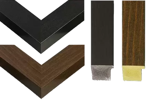
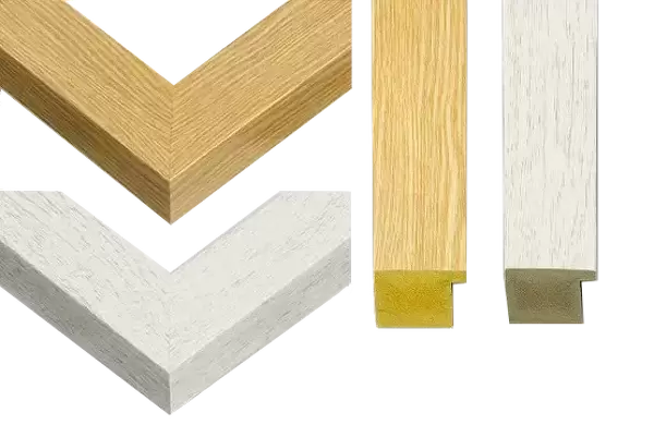
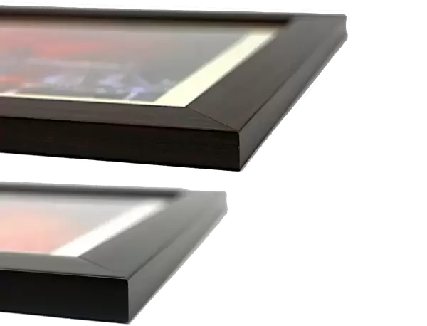
30"x26" (78x68cm) Modern Frame
Discover the wonders of cell biology with our Framed Prints from Media Storehouse. This captivating piece features a stunning immunofluorescent micrograph of a human epithelial cell in the interphase stage of mitosis, captured by Science Photo Library. Each print is meticulously framed in a sleek, modern design, ensuring your scientific masterpiece becomes a focal point in your home or office. Immerse yourself in the intricacies of cell division and elevate your space with this beautiful, high-quality print. Order yours today and bring the magic of science into your world.
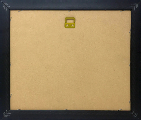
Wood effect frame, card mounted, 24x20 archival quality photo print. Overall outside dimensions 30x26 inches (76x68cm). Environmentally and ozone friendly, 43mm wide x 32mm Polycore® moulding has the look of real wood, is durable and light and easy to hang. Biodegradable and made with non-chlorinated gases (no toxic fumes) it is efficient; producing 100 tons of polystyrene can save 300 tons of trees! Prints are glazed with lightweight, shatterproof, optical clarity acrylic (providing the same general protection from the environment as glass). The back is stapled hardboard with a sawtooth hanger attached. Note: To minimise original artwork cropping, for optimum layout, and to ensure print is secure, the visible print may be marginally smaller
Contemporary Framed and Mounted Prints - Professionally Made and Ready to Hang
Estimated Image Size (if not cropped) is 59.9cm x 59.9cm (23.6" x 23.6")
Estimated Product Size is 68.2cm x 78.2cm (26.9" x 30.8")
These are individually made so all sizes are approximate
Artwork printed orientated as per the preview above, with landscape (horizontal) or portrait (vertical) orientation to match the source image.
FEATURES IN THESE COLLECTIONS
EDITORS COMMENTS
This print showcases the intricate process of cell division in a human epithelial cell during the interphase stage of mitosis. The image, captured using immunofluorescent light microscopy, reveals the remarkable complexity and beauty inherent in our body's cellular machinery. In this snapshot of early mitotic cycle, we witness the chromosomes (depicted in blue) beginning to duplicate and pair up within the parent cell's nucleus. Simultaneously, microtubules (shown in green), like delicate fibers, have formed and are preparing to separate these chromosome pairs to opposite poles of the cell through cytokinesis. The significance of this moment lies in its contribution to our understanding of normal physiology and healthy cellular function. Mitosis is an essential biological process responsible for generating new body cells with identical genetic material. Through meticulous replication and division, two identical daughter cells will emerge from this single entity. Science Photo Library has masterfully captured not only the scientific aspects but also the artistic appeal found within this microscopic world. This mesmerizing composition invites us into a realm where genetics meet fluorescence, cytoplasm meets cytology, and biology intertwines with cellular dynamics. As we gaze upon this extraordinary image print by Science Photo Library, we gain insight into one of nature's most fundamental processes – cell division – reminding us once again that even at such minuscule scales, there exists immense beauty waiting to be discovered.
MADE IN THE UK
Safe Shipping with 30 Day Money Back Guarantee
FREE PERSONALISATION*
We are proud to offer a range of customisation features including Personalised Captions, Color Filters and Picture Zoom Tools
SECURE PAYMENTS
We happily accept a wide range of payment options so you can pay for the things you need in the way that is most convenient for you
* Options may vary by product and licensing agreement. Zoomed Pictures can be adjusted in the Basket.