Acrylic Blox > Popular Themes > Human Body
Acrylic Blox : MRI brain scan F006 / 9208


![]()
Mounted Prints from Science Photo Library
MRI brain scan F006 / 9208
Brain scan. False-colour magnetic resonance imaging (MRI) scan of a human head containing a healthy brain, seen in horizontal view. At upper frame, the two eyeballs can be seen. Between the eyes is the nasal cavity containing the concha, thin scroll- like bones. MRI scanning is a diagnostic technique in which a powerful electromagnet and radio waves create cross-section images of body regions
Science Photo Library features Science and Medical images including photos and illustrations
Media ID 9252909
© PASIEKA/SCIENCE PHOTO LIBRARY
Axial Section Brain Anatomy Brain Scan Cerebrum Cross Section Eyeballs Hemispheres Human Brain Human Head Magnetic Resonance Image Magnetic Resonance Imaging Mri Scan Scientific Imaging Brain Cutouts Nervous System
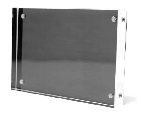
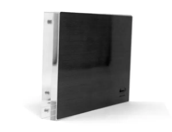
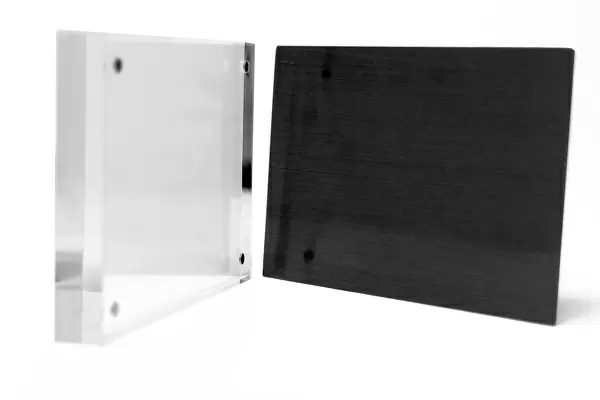
8"x6" (20x15cm) Acrylic Blox
Your photographic print is held in place by magnets and a micro thin sheet of metal covering the back of a 20mm piece of clear acrylic. Your print is held in place with magnets so can easily be replaced if needed.
Streamlined, one sided modern and attractive table top print
Estimated Product Size is 15.2cm x 20.3cm (6" x 8")
These are individually made so all sizes are approximate
Artwork printed orientated as per the preview above, with portrait (vertical) orientation to match the source image.
EDITORS COMMENTS
This print showcases an intricate MRI brain scan, labeled as F006 / 9208. The image displays a false-color representation of a healthy human brain in a horizontal view. Against a pristine white background, the complexity and beauty of our most vital organ are revealed. At the upper frame of the scan, we can observe two eyeballs, reminding us of their close connection to our neurological system. Between these eyes lies the nasal cavity, housing delicate concha bones that resemble elegant scrolls. This comprehensive magnetic resonance imaging (MRI) technique utilizes powerful electromagnets and radio waves to generate cross-sectional images of various body regions. The significance of this diagnostic tool cannot be overstated; it allows medical professionals to delve into the inner workings of our bodies with unprecedented precision. In this particular case, we witness an axial section through the brain, providing insight into its structure and functionality. The cerebral hemispheres take center stage in this composition - they are responsible for higher cognitive functions such as memory, language processing, and problem-solving. As we marvel at this medical illustration's scientific imaging prowess, let us appreciate how MRI scans contribute immensely to healthcare by aiding in accurate diagnoses and treatment plans. This remarkable photograph is part of PASIEKA/SCIENCE PHOTO LIBRARY's collection – an invaluable resource for those seeking knowledge about anatomy or conducting research within the medical field.
MADE IN THE UK
Safe Shipping with 30 Day Money Back Guarantee
FREE PERSONALISATION*
We are proud to offer a range of customisation features including Personalised Captions, Color Filters and Picture Zoom Tools
SECURE PAYMENTS
We happily accept a wide range of payment options so you can pay for the things you need in the way that is most convenient for you
* Options may vary by product and licensing agreement. Zoomed Pictures can be adjusted in the Basket.