Fraud Blocker Skip to main content

Brain Anatomy Collection

"Unveiling the Intricacies

Background imageBrain Anatomy Collection: MRI brain scan F006 / 9208

MRI brain scan F006 / 9208
Brain scan. False-colour magnetic resonance imaging (MRI) scan of a human head containing a healthy brain, seen in horizontal view. At upper frame, the two eyeballs can be seen

Background imageBrain Anatomy Collection: Coronal slice through a healthy human brain

Coronal slice through a healthy human brain
Human brain. Vertical slice through a healthy brain, sectioned coronally from left to right. This cut in the middle of the brain exposes major anatomical regions

Background imageBrain Anatomy Collection: Coloured MRI scan of the human head F007 / 4202

Coloured MRI scan of the human head F007 / 4202
Brain scan. False-colour magnetic resonance imaging (MRI) scan of a human head containing a healthy brain, seen in horizontal view. At upper frame, the two eyeballs can be seen

Background imageBrain Anatomy Collection: Coloured MRI scan of the human head F007 / 4205

Coloured MRI scan of the human head F007 / 4205
Brain scan. False-colour magnetic resonance imaging (MRI) scan of a human head containing a healthy brain, seen in horizontal view. At upper frame, the two eyeballs can be seen

Background imageBrain Anatomy Collection: Coloured MRI scan of the human head F006 / 9210

Coloured MRI scan of the human head F006 / 9210
Brain scan. Computer enhanced Magnetic Resonance Imaging (MRI) scan through a human head, showing a healthy brain in side view. The face is seen in profile at left

Background imageBrain Anatomy Collection: Coloured MRI scan of the human head F006 / 9205

Coloured MRI scan of the human head F006 / 9205
Brain scan. False-colour magnetic resonance imaging (MRI) scan of a human head containing a healthy brain, seen in horizontal view. At upper frame, the two eyeballs can be seen

Background imageBrain Anatomy Collection: Coloured MRI scan of the human head F006 / 9204

Coloured MRI scan of the human head F006 / 9204
Brain scan. Computer enhanced Magnetic Resonance Imaging (MRI) scan through a human head, showing a healthy brain in side view. The face is seen in profile at left

Background imageBrain Anatomy Collection: Coloured MRI scan of the human head F006 / 9200

Coloured MRI scan of the human head F006 / 9200
Brain scan. Computer enhanced Magnetic Resonance Imaging (MRI) scan through a human head, showing a healthy brain in side view. The face is seen in profile at left

Background imageBrain Anatomy Collection: Human head, MRI F006 / 9197

Human head, MRI F006 / 9197
Brain scan. Computer enhanced Magnetic Resonance Imaging (MRI) scan through a human head, showing a healthy brain in side view. The face is seen in profile at left

Background imageBrain Anatomy Collection: Coloured MRI scan of the human head F006 / 9191

Coloured MRI scan of the human head F006 / 9191
Brain scan. Computer enhanced Magnetic Resonance Imaging (MRI) scan through a human head, showing a healthy brain in side view. The face is seen in profile at left

Background imageBrain Anatomy Collection: Coloured MRI scan of the human head F006 / 9192

Coloured MRI scan of the human head F006 / 9192
Brain scan. Computer enhanced Magnetic Resonance Imaging (MRI) scan through a human head, showing a healthy brain in side view. The face is seen in profile at left

Background imageBrain Anatomy Collection: Abstract coloured MRI scan of the human brain

Abstract coloured MRI scan of the human brain
Brain scan abstract. Coloured Magnetic Resonance Imaging (MRI) scan through a human head, showing a healthy brain in side view. Radiating light is emanating from the background

Background imageBrain Anatomy Collection: Computer artwork of the human brain (top view)

Computer artwork of the human brain (top view)
Human brain. Computer artwork of the human brain, seen in top view. Surface folds and convolutions of the left and right cerebral hemispheres are seen

Background imageBrain Anatomy Collection: Brain and vision

Brain and vision
Illustration of visual pathways from the retina of the eye to the brain cortex (seen from below). At the back of each eye (at top)

Background imageBrain Anatomy Collection: Brain superimposed on colour X-ray of human skull

Brain superimposed on colour X-ray of human skull
Healthy brain in skull. Coloured computer-enhanced X-ray of the human skull in side view, with a healthy brain superimposed on it. The brain fits tightly into, and is protected by, the skull

Background imageBrain Anatomy Collection: Graphic of brain in head, with concentric circles

Graphic of brain in head, with concentric circles
Human brain. Computer graphic of a side view of a male head with mesh contours, and a human brain superimposed. Concentric circles are emanating from the temporal region of the brain

Background imageBrain Anatomy Collection: Coloured MRI scan of the human head (side view)

Coloured MRI scan of the human head (side view)
Brain scan. Coloured Magnetic Resonance Imaging (MRI) scan through a human head, showing a healthy brain in side view. The face is seen in profile at left

Background imageBrain Anatomy Collection: Computer-enhanced MRI brain scan (side view)

Computer-enhanced MRI brain scan (side view)
Healthy brain. Computer-enhanced Magnetic Reson- ance Imaging (MRI) scan of a human brain, in side view. This is a sagittal " slice" through the brain in profile

Background imageBrain Anatomy Collection: Brain scan superimposed on contour of human head

Brain scan superimposed on contour of human head

Background imageBrain Anatomy Collection: Coloured artwork of the human brain (side view)

Coloured artwork of the human brain (side view)
Brain. Computer-enhanced historical illustration of the sectioned human brain with cranial nerves, seen in side view. At top are surface folds of the cerebrum

Background imageBrain Anatomy Collection: Mascagni artwork of human brain & blood vessels

Mascagni artwork of human brain & blood vessels
Human Brain. Computer-enhanced historical artwork of the blood supply to the top part of a healthy human brain. The top of the two cerebral hemispheres of the brain is seen with the eyes

Background imageBrain Anatomy Collection: Coloured illustration of sectioned brain in head

Coloured illustration of sectioned brain in head
Brain. Computer-coloured illustration of a human head in side view, sectioned to show the brain and its blood vessels. The face is at right; cavities (pink) of the nose, mouth and pharynx are seen

Background imageBrain Anatomy Collection: Anatomy of head

Anatomy of head
Brain. Computer-coloured illustration of a human head in side view, sectioned to show the brain and its blood vessels. The face is at right; cavities (red) of the nose, mouth and pharynx are seen

Background imageBrain Anatomy Collection: Visible Human Project: sectioned male head

Visible Human Project: sectioned male head

Background imageBrain Anatomy Collection: Artwork of the human brain in head, profile

Artwork of the human brain in head, profile
Human brain. Illustration of a normal brain inside a human head shown in profile. The folded area on top, which occupies most of the brain, is the cerebrum

Background imageBrain Anatomy Collection: Sagittal slice through a healthy human brain

Sagittal slice through a healthy human brain
Human brain. Slice through a healthy brain, sectioned sagittally in the midline from front (at left) to back. This longitudinal cut exposes major regions of the brain

Background imageBrain Anatomy Collection: Artwork: brain frontal areas that control the body

Artwork: brain frontal areas that control the body
Brain-body control. Illustration of a section through the cortex of the frontal lobe, showing brain areas that control voluntary movements in parts of the body

Background imageBrain Anatomy Collection: Illustration of the whole brain in the human head

Illustration of the whole brain in the human head. This side view shows the extensive convolutions of the cerebrum (white); with the cerebellum below it, here coloured red

Background imageBrain Anatomy Collection: Illustration of human muscle and movement systems

Illustration of human muscle and movement systems
Muscle and movement. Illustration of human muscle anatomy and movement systems. Movement depends on the interaction of multiple systems in the body, including muscle, bones and joints

Background imageBrain Anatomy Collection: Coloured X-ray of human skull, brain superimposed

Coloured X-ray of human skull, brain superimposed
Human skull with brain. Coloured computer-enhanced X-ray of the human skull in side view, with a healthy brain superimposed on it

Background imageBrain Anatomy Collection: Mascagni artwork of human brain

Mascagni artwork of human brain
Human Brain. Computer-enhanced historical artwork of the human brain. The bottom of the brain is seen here with the forebrain at top of image

Background imageBrain Anatomy Collection: Short-term memory mechanisms, artwork

Short-term memory mechanisms, artwork
Short-term memory mechanisms. Artwork showing the neural signals (yellow arrows) in short-term memory storage. The signals travel between the eyes, the thalamus (orange), the hippocampus (red)


All Professionally Made to Order for Quick Shipping

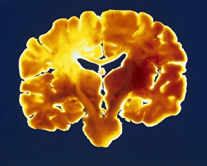
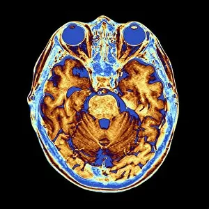
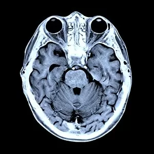
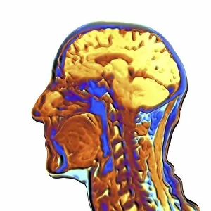
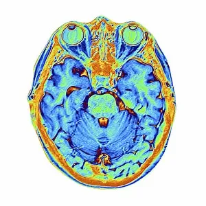
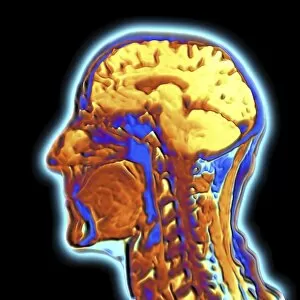
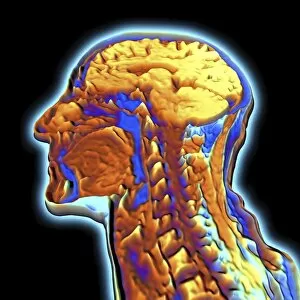
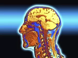
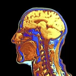
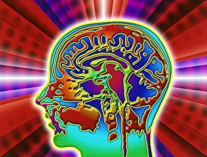
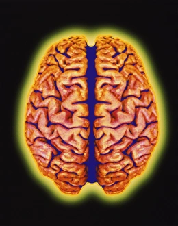
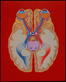
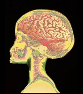
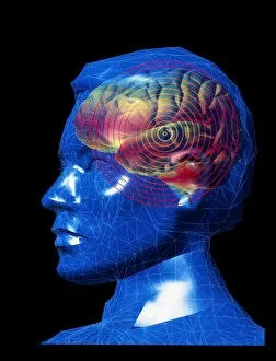
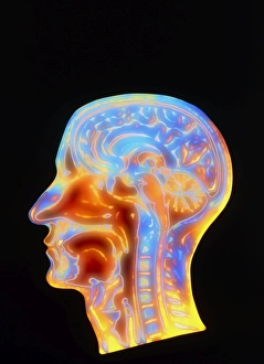
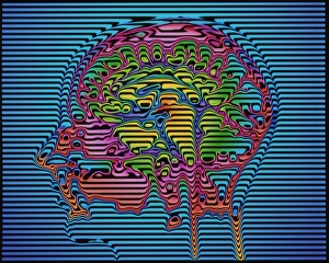
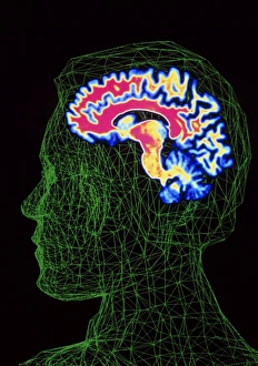
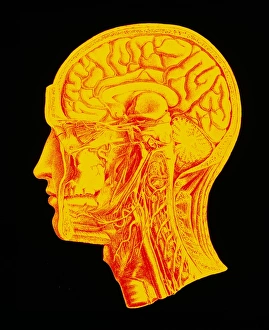
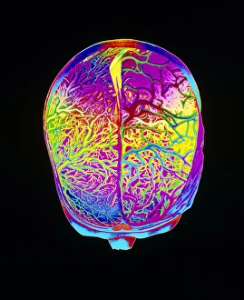
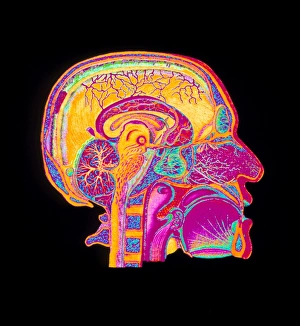
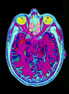
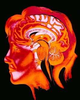
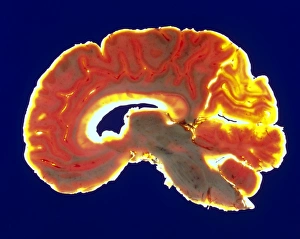
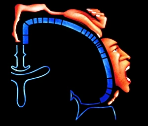
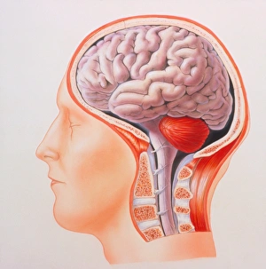
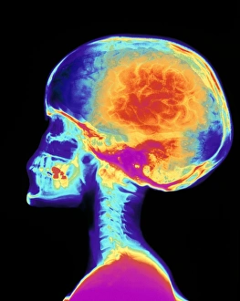
"Unveiling the Intricacies: Exploring Brain Anatomy through Coloured MRI Scans" Step into the fascinating world as we delve deep into the intricate structures that make up this remarkable organ. Through coloured MRI scans, we gain a captivating side view of the human head, revealing a mesmerizing tapestry of neural pathways and cerebral wonders. In one scan, labeled as MRI brain scan F006 / 9208, we witness a coronal slice through a healthy human brain. The vibrant hues highlight distinct regions such as the frontal lobe responsible for decision-making and problem-solving, while the parietal lobe governs sensory perception and spatial awareness. Moving on to another captivating image—coloured MRI scan F007 / 4202—we are presented with an awe-inspiring display of interconnectedness. Neurons weave their way through convoluted folds known as gyri and grooves called sulci, forming an intricate network crucial for information processing. As our exploration continues, coloured MRI scans like F007 / 4205 and F006 / 9210 offer glimpses into specific areas within this complex organ. We observe the temporal lobe housing auditory processing centers where sounds transform into meaningful language or music that resonates within us deeply. The journey takes us further inside with images like F006 / 9205 and F006 / 9204 unveiling deeper layers of understanding. Here lies the limbic system—a nexus of emotions—where joy intertwines with sorrow in delicate balance orchestrated by structures like amygdala and hippocampus. With each new scan - be it F006/9200 or even Human head, MRI F006/9197 - we uncover more secrets hidden beneath our skull's protective shield. These snapshots reveal how different lobes collaborate seamlessly to shape our thoughts, memories, movements; they showcase how intricately intertwined systems work harmoniously to maintain homeostasis.