Home > Popular Themes > Human Body
Oocyte, light micrograph

![]()
Wall Art and Photo Gifts from Science Photo Library
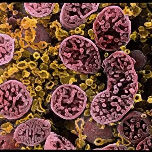
Oocyte, light micrograph
Oocyte. Light micrograph of a section through an oocyte within a early antral follicle in an ovary. Oocytes are immature ova, or egg cells. It has a very large active nucleus (large, round, centre) compared with those seen in the many surrounding granulosa cells of the follicle. Its cytoplasm contains a growing supply of organelles, proteins and messenger RNA required for metabolic and synthetic functions should the oocyte be fertilised and become the activated zygote. The surface of the oocyte is surrounded by a layer of special proteins called the zona pellucida which is an important barrier recognising species-specific sperm and preventing multiple sperm from fertilisation. Magnification: x140 when printed 10 centimetres wide
Science Photo Library features Science and Medical images including photos and illustrations
Media ID 9247507
© MICROSCAPE/SCIENCE PHOTO LIBRARY
Cell Biology Cytological Cytology Egg Cell Female Reproductive System Follicular Gametocyte Germ Cell Histological Histology Immature Nucleus Oocyte Organelle Organelles Ovarian Ovary Ovum Sexual Reproduction Stain Stained Tissue Zona Pellucida Cells Light Micrograph Light Microscope Section Sectioned
EDITORS COMMENTS
This print showcases the intricate details of an oocyte, an immature egg cell found within an early antral follicle in the ovary. The image, captured using a light microscope at a magnification of x140, reveals the remarkable complexity and potential of this tiny reproductive cell. The oocyte's large active nucleus takes center stage in this micrograph, contrasting with the numerous granulosa cells surrounding it. Within its cytoplasm lie a wealth of organelles, proteins, and messenger RNA that are essential for metabolic and synthetic functions if fertilization occurs and it transforms into a zygote. A crucial component protecting the oocyte is its outer layer known as the zona pellucida. Composed of special proteins, this barrier plays a vital role in recognizing species-specific sperm while preventing multiple sperm from fertilizing the egg. This stunning visual representation not only highlights the beauty inherent in biological structures but also emphasizes their significance in sexual reproduction. It provides insight into the complex processes occurring within our bodies on a microscopic level. Displayed by Science Photo Library, this print serves as a reminder of both our awe-inspiring anatomy and ongoing scientific advancements that enable us to explore these intricacies further.
MADE IN THE UK
Safe Shipping with 30 Day Money Back Guarantee
FREE PERSONALISATION*
We are proud to offer a range of customisation features including Personalised Captions, Color Filters and Picture Zoom Tools
SECURE PAYMENTS
We happily accept a wide range of payment options so you can pay for the things you need in the way that is most convenient for you
* Options may vary by product and licensing agreement. Zoomed Pictures can be adjusted in the Basket.