Fraud Blocker Skip to main content

Ovary Collection

The ovary, a vital organ in the female reproductive system, is a fascinating subject of study

Background imageOvary Collection: Coloured SEM of mitochondria in ovarian cells

Coloured SEM of mitochondria in ovarian cells
Mitochondria. Coloured Scanning Electron Micro- graph (SEM) of mitochondria and smooth endoplasmic reticulum in an ovarian granulosa- lutein cell

Background imageOvary Collection: Deer anatomy, artwork

Deer anatomy, artwork
Deer anatomy. Computer artwork showing the internal anatomy of a deer (family Cervidae). Anatomical features shown here include the skeleton, lungs, intestines, bladder

Background imageOvary Collection: Ovarian cancer, light micrograph C015 / 7103

Ovarian cancer, light micrograph C015 / 7103
Ovary cancer. Light micrograph of a section through a Krukenberg tumour (down centre) of the ovary. This is a secondary cancer that spreads (metastasises) from the digestive system

Background imageOvary Collection: Ovarian follicle, SEM

Ovarian follicle, SEM
Ovarian follicle. Coloured scanning electron micrograph (SEM) of a fracture through a secondary follicle in the ovary. The oocyte (developing egg) is orange and its central nucleus is darker orange

Background imageOvary Collection: Whelk, Buccinum undatum 1, sea snails, Cymbium species 2,3, whelk with ovary, Buccinum ampullatum 4

Whelk, Buccinum undatum 1, sea snails, Cymbium species 2,3, whelk with ovary, Buccinum ampullatum 4
FLO4652550 Whelk, Buccinum undatum 1, sea snails, Cymbium species 2,3, whelk with ovary, Buccinum ampullatum 4, shell with egg nest, Vespertilio and Melicera 5, whelk with egg nest

Background imageOvary Collection: Anterior view of a normal uterus with ovaries, fallopian tubes, and broad ligament

Anterior view of a normal uterus with ovaries, fallopian tubes, and broad ligament
Horizontal, Detail, Anatomy, Diagram, Illustration, Medical, Uterus, Biology, Reproduction, Normal, Cut Out, White Background, Artwork, Female Likeness, Color Image, Front View, Close Up

Background imageOvary Collection: Whelks and sea snails with eggs

Whelks and sea snails with eggs
Whelk, Buccinum undatum 1, sea snails, Cymbium species 2, 3, whelk with ovary, Buccinum ampullatum 4, shell with egg nest, Vespertilio and Melicera 5, whelk with egg nest, Buccinum undatum 6

Background imageOvary Collection: The Plague of Crickets in Algeria (engraving)

The Plague of Crickets in Algeria (engraving)
1595535 The Plague of Crickets in Algeria (engraving) by English School, (19th century); Private Collection; (add.info.: The Plague of Crickets in Algeria)

Background imageOvary Collection: Woman standing, body facing forward head turned to one side, illustration overlay showing skeleton

Woman standing, body facing forward head turned to one side, illustration overlay showing skeleton and inner organs

Background imageOvary Collection: Diagram showing the interaction between female sexual organs and the brain, on one side

Diagram showing the interaction between female sexual organs and the brain, on one side, the normal reproductive cycle, and on the other, the effect of the contraceptive pill

Background imageOvary Collection: Cross section illustration of human ovary

Cross section illustration of human ovary

Background imageOvary Collection: 3D rendering of healthy female internal organs

3D rendering of healthy female internal organs

Background imageOvary Collection: Ovarian cyst, endoscope view C017 / 6800

Ovarian cyst, endoscope view C017 / 6800
Ovarian cyst. Endoscope (laparascope) view of a cyst (white) on a patients ovary. Ovarian cysts are common in women of child-bearing age

Background imageOvary Collection: Uterus, endoscope view C017 / 6805

Uterus, endoscope view C017 / 6805
Uterus. Endoscope (laparascope) view of a healthy uterus (womb, large, upper centre), with the two fallopian tubes (left and right) and an ovary (white, lower right)

Background imageOvary Collection: Uterus, Ovary during an ectopic pregnancy, drawing

Uterus, Ovary during an ectopic pregnancy, drawing
Medicine - Anatomy - Obstetrics - Internal female reproductive system. Uterus. Tubes. Ovary during an ectopic pregnancy. Drawing

Background imageOvary Collection: Side view of cross-section model of Puffer Fish with mottled skin camouflage and sharp teeth

Side view of cross-section model of Puffer Fish with mottled skin camouflage and sharp teeth, cross-section of stomach showing deadly toxic liver, ovary and intestines

Background imageOvary Collection: Comparison of healthy and unhealthy female organs

Comparison of healthy and unhealthy female organs
3D rendering comparing healthy female organs and unhealthy female organs

Background imageOvary Collection: 3D rendering of unhealthy female internal organs

3D rendering of unhealthy female internal organs

Background imageOvary Collection: Calcified cysts of the ovary

Calcified cysts of the ovary
Pelvic X-ray (front view) showing calcified cysts in a womans ovary (white, lower left)

Background imageOvary Collection: Menopause, conceptual artwork

Menopause, conceptual artwork. Menopause is the cessation of menstruation. It usually occurs between the ages of 45 and 55

Background imageOvary Collection: Cow anatomy, artwork

Cow anatomy, artwork
Cow anatomy. Computer artwork showing the internal anatomy of a domesticated cow (Bos primigenius). Anatomical features shown here include the skeleton, lungs, intestines, bladder

Background imageOvary Collection: Bletia purpurea

Bletia purpurea - Bletia purpurea. Longidunal and traverse section of the ovary. Watercolour by Franz Bauer, 1801. Paper, watercolour. Botany Library, Bauer Unit, Unfinished Drawings of Plants 3: no

Background imageOvary Collection: Sparmannia discolor by Matilda Smith

Sparmannia discolor by Matilda Smith
Graphite on paper, 1880s by Matilda Smith (1854-1926). Held in the Library and Archives

Background imageOvary Collection: Cross section illustration of internal anatomy of female lizard

Cross section illustration of internal anatomy of female lizard

Background imageOvary Collection: Life cycle of a flowering plant

Life cycle of a flowering plant

Background imageOvary Collection: Digital illustration of human neuroendocrine system in female body

Digital illustration of human neuroendocrine system in female body

Background imageOvary Collection: Illustration of broadleaf flower showing ovules inside the ovary

Illustration of broadleaf flower showing ovules inside the ovary

Background imageOvary Collection: Illustration of fertilization of flower, pollen grain containing male nuclei on stigma

Illustration of fertilization of flower, pollen grain containing male nuclei on stigma, male nucleus travelling down pollen tube, female nuclei inside ovule

Background imageOvary Collection: Cross section biomedical illustration of egg development inside ovary during human menstrual cycle

Cross section biomedical illustration of egg development inside ovary during human menstrual cycle

Background imageOvary Collection: Microscopic cross section of a female ovary

Microscopic cross section of a female ovary

Background imageOvary Collection: Microscopic view of ovules

Microscopic view of ovules


All Professionally Made to Order for Quick Shipping

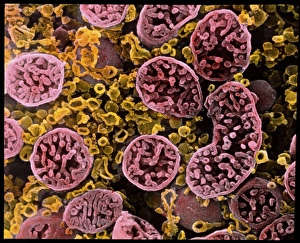
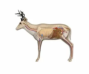
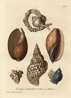
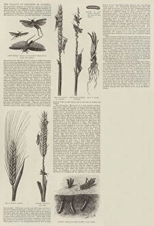
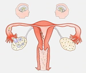
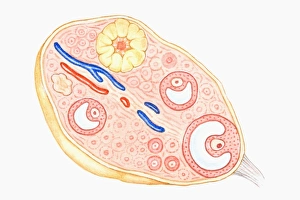
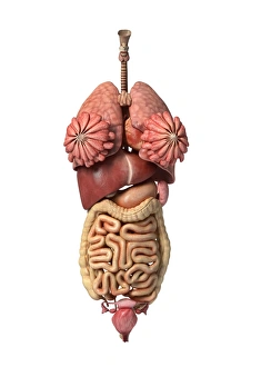
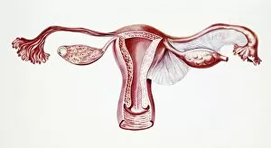
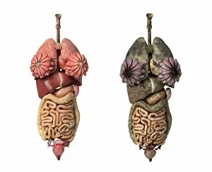
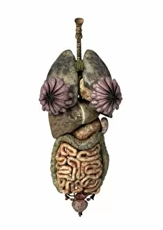
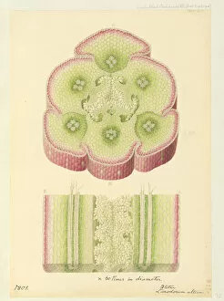
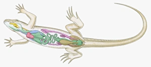
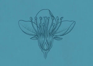
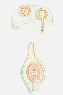
The ovary, a vital organ in the female reproductive system, is a fascinating subject of study. Through advanced imaging techniques, such as coloured scanning electron microscopy (SEM), we can now explore its intricate structures like never before. In one captivating image, we observe mitochondria within ovarian cells under SEM. These tiny powerhouses play a crucial role in cellular energy production and are essential for the proper functioning of the ovary. However, not all aspects of the it can beautiful or benign. Ovarian cancer, depicted in a light micrograph C015/7103, reminds us of the importance of early detection and effective treatments for this devastating disease. On a more positive note, an SEM image showcases an ovarian follicle - an essential structure that nurtures and releases mature eggs during each menstrual cycle. This delicate process is fundamental to female fertility. Moving beyond human ovaries, we discover surprising similarities between different species. The opium poppy's it also reveals itself through SEM imagery – reminding us that nature's wonders extend far beyond our own bodies. A cross-section biomedical illustration provides insight into how the ovary fits within the broader context of the female reproductive system. It highlights its connection to other organs like fallopian tubes and broad ligaments – forming an intricate network responsible for conception and childbirth. Delving into history takes us back centuries ago when Johannes de Ketham's woodcut illustrations showcased pregnant women with their anatomical features meticulously detailed. Such depictions offer glimpses into medical knowledge from bygone eras. Nature continues to captivate with images showcasing whelks and sea snails carrying their precious cargo: eggs. These creatures remind us that reproduction is not exclusive to humans but rather spans across diverse organisms on our planet. Returning to artistry from another era brings forth painted cardboard depicting female genitals from around 1830 – highlighting society's fascination with anatomy throughout history.