Fraud Blocker Skip to main content

Ovary Collection (page 6)

The ovary, a vital organ in the female reproductive system, is a fascinating subject of study

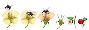
Background imageOvary Collection: Bee pollinating a flower, artwork C018 / 0297

Bee pollinating a flower, artwork C018 / 0297
Bee pollinating a flower. Sequence of artworks showing how a bee (left) lands on a flower to drink nectar (centre left), brushing up against the pollen-bearing anthers

Background imageOvary Collection: Flower structure, artwork C017 / 7232

Flower structure, artwork C017 / 7232
Flower structure. Computer artwork showing the main structures of a typical flower. The flower is the reproductive unit of some angiosperm plants

Background imageOvary Collection: Ovarian cyst in abdomen, artwork

Ovarian cyst in abdomen, artwork
Ovarian cyst in abdomen. Artwork of a large ovarian cyst (centre left) in a dissected human abdomen, seen from above. The uterus (centre) and the ovaries are part of the female reproductive system

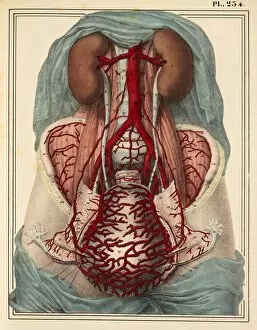
Background imageOvary Collection: Uterine arteries, 1825 artwork

Uterine arteries, 1825 artwork
Uterine arteries. Dissection showing the arteries (red) of the uterus (lower centre) of a woman who died six days after giving birth. The ovaries and fallopian tubes are either side

Background imageOvary Collection: Ectopic pregnancy, artwork C016 / 8737

Ectopic pregnancy, artwork C016 / 8737
Ectopic pregnancy, artwork. This is a pregnancy where the embryo (upper left) implants outside the uterus, usually as here in the Fallopian tube that should channel the fertilised egg to the uterus

Background imageOvary Collection: Ovulation and fertilisation, artwork

Ovulation and fertilisation, artwork
Ovulation and fertilisation. Computer artwork of the female reproductive system, showing the development of an ovum (egg, small circle) inside an ovary (oval)

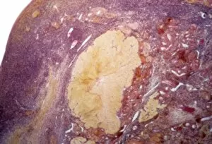
Background imageOvary Collection: Dermoid ovarian cyst C015 / 6910

Dermoid ovarian cyst C015 / 6910
Dermoid ovarian cyst. Light micrograph of a section through an ovary with a dermoid, or teratoma, ovarian cyst. This is a benign (non-cancerous) cyst that arises from germ (sex) cells

Background imageOvary Collection: Ovarian fibroid C015 / 6749

Ovarian fibroid C015 / 6749
Ovarian fibroid. Gross specimen of a sectioned ovarian fibroid. A fibroid is a fibrous benign tumour originating from muscular tissue

Background imageOvary Collection: Ovarian fibroid C015 / 6748

Ovarian fibroid C015 / 6748
Ovarian fibroid. Gross specimen of a sectioned ovarian fibroid. A fibroid is a fibrous benign tumour originating from muscular tissue

Background imageOvary Collection: Dermoid ovarian cyst C015 / 6056

Dermoid ovarian cyst C015 / 6056
Dermoid ovarian cyst. Light micrograph of a section through an ovary with a dermoid, or teratoma, ovarian cyst. This is a benign (non-cancerous) cyst that arises from germ (sex) cells

Background imageOvary Collection: Ovarian cysts, ultrasound scan C017 / 8018

Ovarian cysts, ultrasound scan C017 / 8018
Ovarian cysts. Ultrasound scan of the abdomen of an 85 year old female patient showing a multicystic mass (upper centre) in an ovary

Background imageOvary Collection: Ovarian cysts, ultrasound scan C017 / 8017

Ovarian cysts, ultrasound scan C017 / 8017
Ovarian cysts. Ultrasound scan of the abdomen of an 85 year old female patient showing a multicystic mass (centre) in an ovary

Background imageOvary Collection: Endometrial cyst, ultrasound scan C017 / 8006

Endometrial cyst, ultrasound scan C017 / 8006
Endometrial cyst. Ultrasound of a scan of an endometrial cyst (black) in the ovary of a 41 year old female patient. Endometriosis is a condition where detached parts of the uterus lining (endometrium)

Background imageOvary Collection: Corpus albicans, light micrograph C015 / 6226

Corpus albicans, light micrograph C015 / 6226
Corpus albicans. Light micrograph of a section through an ovary showing a corpus albicans (centre). Each menstrual cycle one egg (ovum) reaches maturity and develops into a Graafian follicle

Background imageOvary Collection: Ovarian fibroid C015 / 6414

Ovarian fibroid C015 / 6414
Ovarian fibroid. Gross specimen of an ovarian fibroid. A fibroid is a fibrous benign tumour originating from muscular tissue

Background imageOvary Collection: Dermoid ovarian cyst C015 / 6402

Dermoid ovarian cyst C015 / 6402
Dermoid ovarian cyst. Light micrograph of a section through an ovary with a dermoid, or teratoma, ovarian cyst. This is a benign (non-cancerous) cyst that arises from germ (sex) cells

Background imageOvary Collection: Prick nose (Lychnis coronaria) C016 / 5687

Prick nose (Lychnis coronaria) C016 / 5687
Prick nose (Lychnis coronaria). Watercolour (Plate 171) by Arthur Harry Church, 12 July 1905

Background imageOvary Collection: Golden columbine (Aquilegia chrysantha) C016 / 5685

Golden columbine (Aquilegia chrysantha) C016 / 5685
Golden columbine (Aquilegia chrysantha). Ink and wash drawing by Arthur Harry Church, 1906. Figure for Types of floral mechanism. Natural History Museum, London, UK

Background imageOvary Collection: Honesty (Lunaria biennis), artwork C016 / 5676

Honesty (Lunaria biennis), artwork C016 / 5676
Hinesty (Lunaria biennis). Watercolour (Plate 146) by Arthur Harry Church. Held in the Botany Library at the Natural History Museum, London, UK

Background imageOvary Collection: Darlingtonia californica, artwork C016 / 5576

Darlingtonia californica, artwork C016 / 5576
California pitcherplant (Darlingtonia californica). Drawing (Plate 134.) by Arthur Harry Church, 1909. Held in the Botany Library at the Natural History Museum, London, UK

Background imageOvary Collection: Hardy larkspur (Delphinium formosum) C016 / 5575

Hardy larkspur (Delphinium formosum) C016 / 5575
Hardy larkspur (Delphinium formosum). Drawing by Arthur Harry Church, 1903. Held in the Botany Library at the Natural History Museum, London, UK

Background imageOvary Collection: Yellow fennel flower (Nigella orientalis) C016 / 5574

Yellow fennel flower (Nigella orientalis) C016 / 5574
Yellow fennel flower (Nigella orientalis). Drawing (Plate 062) by Arthur Harry Church, 1905. Held in the Botany Library at the Natural History Museum, London, UK

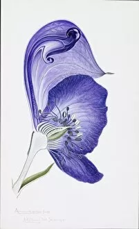
Background imageOvary Collection: Monk s-hood (Aconitumm napellus) C016 / 5572

Monk s-hood (Aconitumm napellus) C016 / 5572
Monk s-hood (Aconitumm napellus). Illustration (Plate 12) by Arthur Harry Church, 1903. Held in the Botany Library at the Natural History Museum, London, UK

Background imageOvary Collection: Flower bud detail, SEM

Flower bud detail, SEM
Detail of inside a flower bud, coloured scanning electron micrograph (SEM). The flower is a cape fuchsia (Phygelius aequalis). Magnification is x14 at an image of 10cm wide

Background imageOvary Collection: Section through a lily ovary with ovules

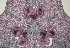
Section through a lily ovary with ovules
Light micrograph of a transverse section through the ovary of a lily, Lilium sp.. Two ovules (female gametes) are visible within each of the three hollow chambers of the ovary

Background imageOvary Collection: Dr. Ephraim McDowell performing the first recorded operation in ovarian surgery in the United

Dr. Ephraim McDowell performing the first recorded operation in ovarian surgery in the United States at Danville
EPHRAIM McDOWELL, 1809. Dr. Ephraim McDowell performing the first recorded operation in ovarian surgery in the United States at Danville, Kentucky, in December 1809. Lithograph, 19th century

Background imageOvary Collection: Liver fluke, transverse section

Liver fluke, transverse section
Liver fluke. Light micrograph of a transverse section through the anterior half of a liver fluke (Fasciola hepatica), a hermaphrodite tremotode worm and endoparasite that infects sheep

Background imageOvary Collection: Nematode worm, transverse sections

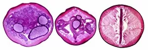
Nematode worm, transverse sections
Nematode worm. Light micrograph of three transverse sections through the body of a female round unsegmented worm (Ascaris lumbricoides)

Background imageOvary Collection: Cubic close-packed crystal structure

Cubic close-packed crystal structure
Haemoglobin molecule. Computer artwork showing the structure of a haemoglobin molecule. Haemoglobin is a metalloprotein that transports oxygen around the body in red blood cells

Background imageOvary Collection: Blood vessels in a frog ovary, SEM

Blood vessels in a frog ovary, SEM
Blood vessels in the ovary of a female frog, coloured scanning electron micrograph (SEM). Frogs produce hundreds of eggs, which are expelled and fertilised in the water

Background imageOvary Collection: Ovulation, artwork

Ovulation, artwork
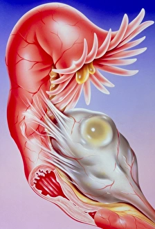
Ovulation. Computer artwork of an egg (yellow) being released from an ovary (bottom), a process known as ovulation. The egg is being released into a fallopian tube (top)

Background imageOvary Collection: False-colour SEM of the ovary surface at ovulation

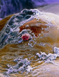
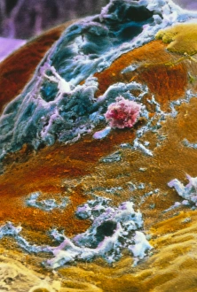
False-colour SEM of the ovary surface at ovulation

Background imageOvary Collection: False-colour SEM of human egg after ovulation

False-colour SEM of human egg after ovulation
Ovarys surface at ovulation. False-colour scanning electron micrograph of the surface of the ovary about one hour after ovulation

Background imageOvary Collection: Ovarian tissue, SEM

Ovarian tissue, SEM
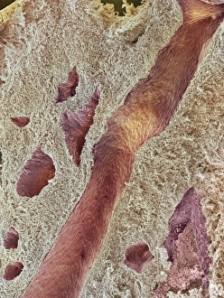
Ovarian tissue. Coloured scanning electron micrograph (SEM) of a section through the medulla of an ovary showing blood vessels (pink)

Background imageOvary Collection: Corpus luteum, SEM

Corpus luteum, SEM
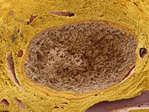
Corpus luteum. Coloured scanning electron micrograph (SEM) of a corpus luteum, or yellow body (brown), in an ovary. The corpus luteum is formed after ovulation
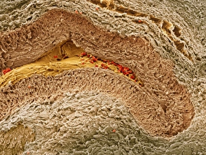
Background imageOvary Collection: Collapsed ovarian follicle, SEM

Collapsed ovarian follicle, SEM
Collapsed ovarian follicle. Coloured scanning electron micrograph (SEM) of an ovarian follicle just after ovulation (release of an egg)

Background imageOvary Collection: Follicles in a human ovary, micrograph

Follicles in a human ovary, micrograph
Developing follicles in a human ovary, light micrograph. Three developing Graafian follicles are seen. A Graafian follicle is a structure in the ovaries from which an ovum (egg cell) is released

Background imageOvary Collection: Artwork of ovum (egg) development in womans ovary

Artwork of ovum (egg) development in womans ovary
Ovum development. Illustration showing the development of a human ovum (egg) in an ovary (pink, at lower centre) during the menstrual cycle

Background imageOvary Collection: Graafian follicles, light micrograph

Graafian follicles, light micrograph
Developing Graafian follicles. Light micrograph of a sectioned human ovary, showing two developing Graafian follicles. A Graafian follicle is a structure in the ovaries from which an ovum (egg cell)

Background imageOvary Collection: Ovarian blood vessel, SEM

Ovarian blood vessel, SEM
Ovarian blood vessel. Coloured scanning electron micrograph (SEM) of a section through a blood vessel in an ovary. Red blood cells are seen in the lumen, which is lined with endothelial cells (brown)

Background imageOvary Collection: Ovarian follicles, light micrograph

Ovarian follicles, light micrograph
Ovarian follicles. Coloured light micrograph of a section through an ovary (orange), showing three primary follicles (pink)
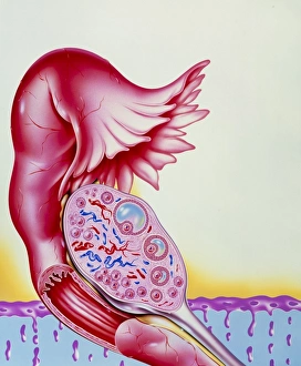
Background imageOvary Collection: Ovarian artery, SEM

Ovarian artery, SEM
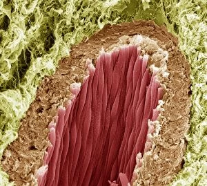
Ovarian artery. Coloured scanning electron micrograph (SEM) of an ovarian artery that has been freeze-fractured to show internal details

Background imageOvary Collection: Illustration of fallopian tube infertility

Illustration of fallopian tube infertility. A fallopian tube extends from the uterus to each ovary; it transports eggs and sperm, and is the site of fertilization


All Professionally Made to Order for Quick Shipping

The ovary, a vital organ in the female reproductive system, is a fascinating subject of study. Through advanced imaging techniques, such as coloured scanning electron microscopy (SEM), we can now explore its intricate structures like never before. In one captivating image, we observe mitochondria within ovarian cells under SEM. These tiny powerhouses play a crucial role in cellular energy production and are essential for the proper functioning of the ovary. However, not all aspects of the it can beautiful or benign. Ovarian cancer, depicted in a light micrograph C015/7103, reminds us of the importance of early detection and effective treatments for this devastating disease. On a more positive note, an SEM image showcases an ovarian follicle - an essential structure that nurtures and releases mature eggs during each menstrual cycle. This delicate process is fundamental to female fertility. Moving beyond human ovaries, we discover surprising similarities between different species. The opium poppy's it also reveals itself through SEM imagery – reminding us that nature's wonders extend far beyond our own bodies. A cross-section biomedical illustration provides insight into how the ovary fits within the broader context of the female reproductive system. It highlights its connection to other organs like fallopian tubes and broad ligaments – forming an intricate network responsible for conception and childbirth. Delving into history takes us back centuries ago when Johannes de Ketham's woodcut illustrations showcased pregnant women with their anatomical features meticulously detailed. Such depictions offer glimpses into medical knowledge from bygone eras. Nature continues to captivate with images showcasing whelks and sea snails carrying their precious cargo: eggs. These creatures remind us that reproduction is not exclusive to humans but rather spans across diverse organisms on our planet. Returning to artistry from another era brings forth painted cardboard depicting female genitals from around 1830 – highlighting society's fascination with anatomy throughout history.