Premium Framed Print : Stroke. Coloured computed tomography (CT) brain scan showing a cerebrovascular accident
![]()

Framed Photos from Science Photo Library
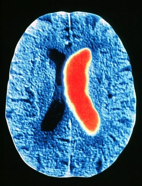
Stroke. Coloured computed tomography (CT) brain scan showing a cerebrovascular accident
Stroke. Coloured computed tomography (CT) brain scan showing a cerebrovascular accident (CVA), or stroke. The red region is an area of internal bleeding, or haemorrhage. Stroke is brain damage caused by the interruption of the brains blood supply or by the leakage of blood through blood vessel walls. The two main causes are high blood pressure and atherosclerosis (narrowing of the arteries due to fat deposition). Strokes vary in severity depending on the site and extent of damage, but can result in long-term paralysis, coma and death. A CT scan is a cross-sectional map of the body created by the computation of X-rays passed through the body at different angles
Science Photo Library features Science and Medical images including photos and illustrations
Media ID 6421953
© PASIEKA/SCIENCE PHOTO LIBRARY
Axial Section Brain Damage Cerebrovascular Accident Circle Circles Computed Tomography Ct Scan Haemorrhage Hemisphere Hemispheres Round Shape Rounded Circular Stroke Vascular Condition Disorder Health Care
17"x15" (43x38cm) Premium Frame
FSC real wood frame with double mounted 10x8 print. Double mounted with white conservation mountboard. Frame moulding comprises stained composite natural wood veneers (Finger Jointed Pine) 39mm wide by 21mm thick. Archival quality Fujifilm CA photo paper mounted onto 1mm card. Overall outside dimensions are 17x15 inches (431x381mm). Rear features Framing tape to cover staples, 50mm Hanger plate, cork bumpers. Glazed with durable thick 2mm Acrylic to provide a virtually unbreakable glass-like finish. Acrylic Glass is far safer, more flexible and much lighter than typical mineral glass. Moreover, its higher translucency makes it a perfect carrier for photo prints. Acrylic allows a little more light to penetrate the surface than conventional glass and absorbs UV rays so that the image and the picture quality doesn't suffer under direct sunlight even after many years. Easily cleaned with a damp cloth. Please note that, to prevent the paper falling through the mount window and to prevent cropping of the original artwork, the visible print may be slightly smaller to allow the paper to be securely attached to the mount without any white edging showing and to match the aspect ratio of the original artwork.
FSC Real Wood Frame and Double Mounted with White Conservation Mountboard - Professionally Made and Ready to Hang
Estimated Image Size (if not cropped) is 18.7cm x 24.4cm (7.4" x 9.6")
Estimated Product Size is 38.1cm x 43.1cm (15" x 17")
These are individually made so all sizes are approximate
Artwork printed orientated as per the preview above, with portrait (vertical) orientation to match the source image.
EDITORS COMMENTS
This print from Science Photo Library showcases a coloured computed tomography (CT) brain scan, revealing the devastating effects of a cerebrovascular accident (CVA), commonly known as a stroke. The vibrant red region within the image represents an area of internal bleeding or haemorrhage, highlighting the severity of this condition. A stroke occurs when there is an interruption in blood supply to the brain or when blood leaks through vessel walls, resulting in significant brain damage. The two primary causes are high blood pressure and atherosclerosis, which refers to the narrowing of arteries due to fat deposition. The impact of strokes can vary depending on their location and extent. They can lead to long-term paralysis, coma, and even death. This image serves as a powerful reminder of the potential consequences associated with this medical condition. Utilizing computed tomography technology, this cross-sectional map provides valuable insights into the intricate details of our body's inner workings. By capturing X-rays at different angles and computing them together, CT scans offer invaluable diagnostic information for healthcare professionals. Science Photo Library has expertly captured this visually striking representation that encompasses various aspects such as disease, health care, medicine, vascular issues while showcasing cut-outs and rounded circular shapes found within our hemispheres. It is important to note that this caption does not mention any commercial use but rather focuses on appreciating the scientific value behind such imagery provided by Science Photo Library.
MADE IN THE UK
Safe Shipping with 30 Day Money Back Guarantee
FREE PERSONALISATION*
We are proud to offer a range of customisation features including Personalised Captions, Color Filters and Picture Zoom Tools
SECURE PAYMENTS
We happily accept a wide range of payment options so you can pay for the things you need in the way that is most convenient for you
* Options may vary by product and licensing agreement. Zoomed Pictures can be adjusted in the Basket.


