Poster Print > Science > SEM
Poster Print : Coloured SEM of mitochondria in ovarian cells


![]()
Poster Prints from Science Photo Library
Coloured SEM of mitochondria in ovarian cells
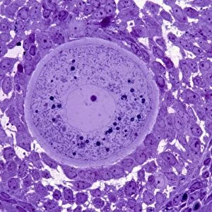
Mitochondria. Coloured Scanning Electron Micro- graph (SEM) of mitochondria and smooth endoplasmic reticulum in an ovarian granulosa- lutein cell. The cell occurs in the corpus luteum of the ovary, which develops after ovulation and secretes the hormone progesterone. Mitochondria (pink) are seen sectioned, containing tubular cristae. Active secretory cells have many mitochondria which play a role in cell respiration. Membranes of smooth endoplasmic reticulum (ER, yellow) are also seen; commonly found in secretory cells, ER synthesizes lipids and transports substances around the cell. Magnification: x21, 300 at 6x7cm size. Magnification: x27, 400 at 4x5 inch size
Science Photo Library features Science and Medical images including photos and illustrations
Media ID 6402825
© PROFESSORS P.M. MOTTA, S. MAKABE & T. NAGURO/SCIENCE PHOTO LIBRARY
Cell Structure Corpus Luteum Cytology Endoplasmic Reticulum Hormone Mitochondria Ovary Progesterone Smooth Micro Biology
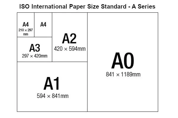
A2 (59.4 x 42cm) Poster Print
"Discover the intricacies of cellular structures with our stunning range of Poster Prints from Media Storehouse. This captivating image showcases a Coloured Scanning Electron Micrograph (SEM) of mitochondria and smooth endoplasmic reticulum in an ovarian granulosa-lutein cell, captured by Science Photo Library. Delve deep into the world of science and bring the beauty of cellular structures into your home or office space. Our high-quality posters are printed on premium paper, ensuring vibrant colours and long-lasting durability. Order now and ignite your curiosity!"
A2 Poster (59.4 x 42cm, 23.4" x 16.5" inches) printed on 170gsm Satin Poster Paper. Securely packaged, rolled and inserted into a strong mailing tube and shipped tracked. Poster Prints are of comparable archival quality to our Photographic prints, they are simply printed on thinner Poster Paper. Whilst we only use Photographic Prints in our frames, you can frame Poster Prints if they are carefully supported to prevent sagging over time.
Poster prints are budget friendly enlarged prints in standard poster paper sizes (A0, A1, A2, A3 etc). Whilst poster paper is sometimes thinner and less durable than our other paper types, they are still ok for framing and should last many years. Our Archival Quality Photo Prints and Fine Art Paper Prints are printed on higher quality paper and the choice of which largely depends on your budget.
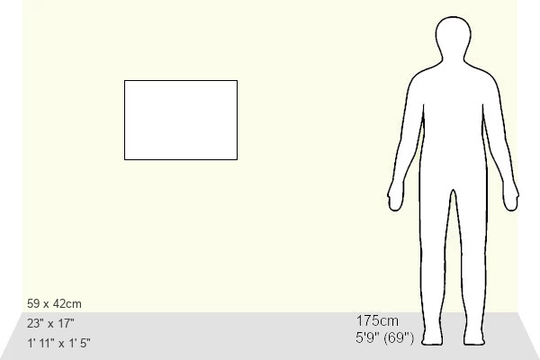
Estimated Image Size (if not cropped) is 51.7cm x 42cm (20.4" x 16.5")
Estimated Product Size is 59.4cm x 42cm (23.4" x 16.5")
These are individually made so all sizes are approximate
Artwork printed orientated as per the preview above, with landscape (horizontal) orientation to match the source image.
EDITORS COMMENTS
This print showcases the intricate beauty of ovarian cells, specifically highlighting mitochondria and smooth endoplasmic reticulum. The image reveals a coloured scanning electron micrograph (SEM) that provides a detailed view of these cellular structures within an ovarian granulosa-lutein cell found in the corpus luteum. Mitochondria, depicted in delicate shades of pink, are prominently displayed with their tubular cristae clearly visible. These energy-producing powerhouses are crucial for cell respiration and play a vital role in active secretory cells like this one. Meanwhile, the yellow membranes represent the smooth endoplasmic reticulum (ER), which is responsible for lipid synthesis and intracellular transportation. The corpus luteum itself forms after ovulation and serves as a site for progesterone secretion – an essential hormone involved in reproductive processes. This image offers valuable insights into the structure and function of these fascinating cells found within the ovary tissue. With magnifications ranging from x21,300 at 6x7cm size to x27,400 at 4x5 inch size, this photograph captures even the tiniest details with remarkable clarity. It serves as a testament to both the complexity of biological systems and the incredible advancements made possible through microscopy techniques. This stunning visual representation was captured by Science Photo Library's team of experts dedicated to showcasing scientific wonders while contributing to our understanding of biology on a microscopic level.
MADE IN THE UK
Safe Shipping with 30 Day Money Back Guarantee
FREE PERSONALISATION*
We are proud to offer a range of customisation features including Personalised Captions, Color Filters and Picture Zoom Tools
SECURE PAYMENTS
We happily accept a wide range of payment options so you can pay for the things you need in the way that is most convenient for you
* Options may vary by product and licensing agreement. Zoomed Pictures can be adjusted in the Basket.