
Cell Structure Collection
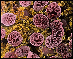
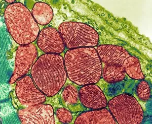
"Exploring the Intricate World of Cell Structure: Unveiling the Wonders Within" Coloured SEM of mitochondria in ovarian cells


All Professionally Made to Order for Quick Shipping
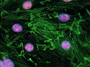
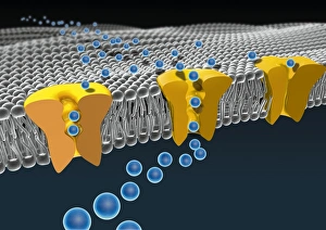
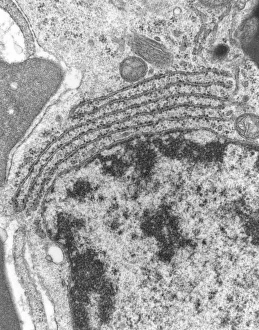
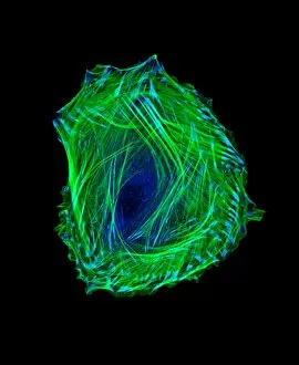
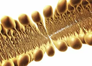
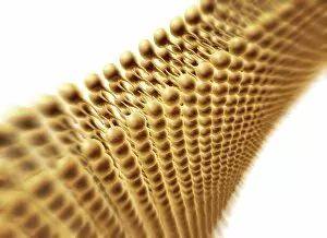
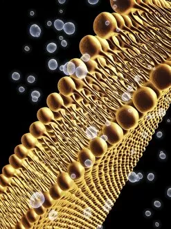
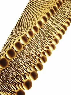
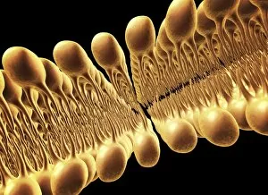
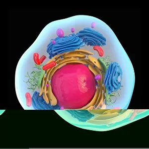
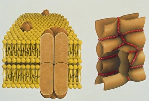
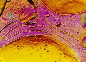
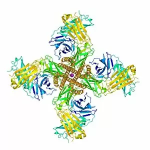
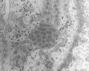
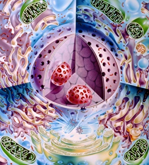
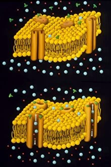
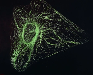
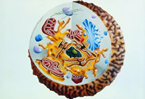
"Exploring the Intricate World of Cell Structure: Unveiling the Wonders Within" Coloured SEM of mitochondria in ovarian cells: Witness the vibrant beauty and complexity of mitochondria, the powerhouses within our cells. Cell membrane, artwork C013 / 7467: Delve into the intricate structure of the cell membrane, a crucial barrier that protects and regulates cellular processes. Mitochondrion, TEM: Peer through the lens of a transmission electron microscope to uncover the detailed architecture of this vital organelle responsible for energy production. Immunofluorescent LM of fibroblast cell nuclei: Illuminate your understanding as fluorescent markers reveal fibroblast cell nuclei, shedding light on their role in tissue repair and maintenance. Animal cell structure: Embark on an exploration into animal cell structure – a complex network comprising various organelles working harmoniously to sustain life. Cell membrane lipid bilayer, artwork F007 / 1477: Dive deep into this artistic representation showcasing how lipids arrange themselves to form a dynamic double-layered boundary around cells. Cell membrane ion channels, artwork C016 / 7689: Discover how specialized proteins create pathways known as ion channels across cell membranes facilitating communication and transport between cells. Sindbis virus capsid protein: Unlocking secrets at a molecular level, witness the captivating structure of Sindbis virus capsid protein – an essential component for viral replication within host cells. Mitochondria, TEM: Explore further with transmission electron microscopy as it reveals intricate details about these energy-producing organelles found in abundance throughout our bodies' cells. Bacillus bacterial genus: Journey into microbial realms by exploring Bacillus bacterial genus - unravelling their unique cellular structures that enable them to thrive in diverse environments. Conceptual image of centriole (s).