Metal Print > Science > SEM
Metal Print : Cartilage cell, SEM


![]()
Metal Prints from Science Photo Library
Cartilage cell, SEM
Cartilage cell. Coloured scanning electron micrograph (SEM) of a section through a chondrocyte cell (centre) in cartilage. Cartilage is a type of connective tissue formed of chondrocyte cells embedded in a matrix (green) of collagen fibres and proteoglycans. The chondrocyte has a large nucleus (pink). The space in the matrix occupied by a chondrocyte cell is called the lacuna. Cartilage provides support for the walls of the airway, and caps the ends of long bones. Magnification: x3150 when printed 10cm wide
Science Photo Library features Science and Medical images including photos and illustrations
Media ID 6420470
© STEVE GSCHMEISSNER/SCIENCE PHOTO LIBRARY
Cartilage Chondrocyte Connective Tissue Horizontal Intercellular Lacuna Material Matrix Nucleus Structural Contents
15"x10" (38x25cm) Metal Print
Discover the intricacy of life with Media Storehouse's Metal Prints. Feast your eyes on this captivating Coloured Scanning Electron Micrograph (SEM) of a chondrocyte cell, the star player in cartilage formation. Each print in our range is meticulously crafted on high-quality metal, ensuring a vibrant and long-lasting display of this mesmerizing Cartilage Cell image from Science Photo Library. Bring the wonders of science into your home or office with Media Storehouse Metal Prints.
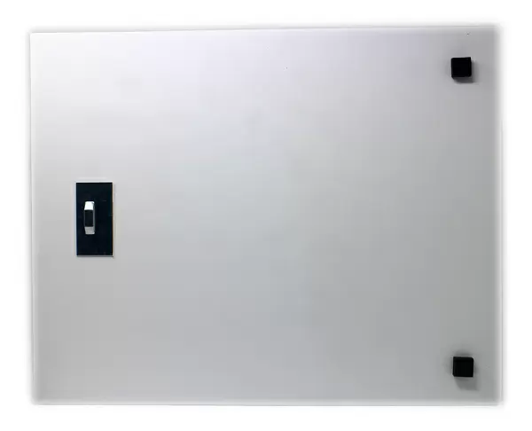
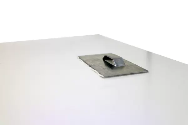
Your image is printed photographically and bonded to a 3.5mm thick, Dibond board (black polyethylene sandwiched between two sheets of white coated aluminium). The panel is then sealed with a gloss protective covering. Supplied complete with a wall mount which holds the print 10mm from the wall.
Made with durable metal and luxurious printing techniques, metal prints bring images to life and add a modern touch to any space
Estimated Product Size is 38.1cm x 25.4cm (15" x 10")
These are individually made so all sizes are approximate
Artwork printed orientated as per the preview above, with landscape (horizontal) or portrait (vertical) orientation to match the source image.
EDITORS COMMENTS
This print showcases the intricate beauty of a cartilage cell, as seen through a scanning electron microscope (SEM). The image reveals a section through a chondrocyte cell at its center, surrounded by the matrix of collagen fibers and proteoglycans that make up cartilage. This connective tissue plays an essential role in providing support for the airway walls and capping the ends of long bones. The vibrant colors highlight different components of this microscopic world. The chondrocyte's large nucleus stands out in striking pink against the green backdrop of the collagen fiber matrix. Within this matrix lies the lacuna, which represents the space occupied by each individual chondrocyte cell. This photograph not only captures structural details but also offers insight into our body's anatomy on a cellular level. It serves as a reminder of how complex and fascinating our own biology can be when viewed from such close proximity. Printed at 10cm wide with an impressive magnification factor of x3150, this image allows us to appreciate both its artistic qualities and scientific significance. Whether you are fascinated by cellular structures or simply intrigued by nature's hidden wonders, this print is sure to captivate your imagination.
MADE IN THE UK
Safe Shipping with 30 Day Money Back Guarantee
FREE PERSONALISATION*
We are proud to offer a range of customisation features including Personalised Captions, Color Filters and Picture Zoom Tools
SECURE PAYMENTS
We happily accept a wide range of payment options so you can pay for the things you need in the way that is most convenient for you
* Options may vary by product and licensing agreement. Zoomed Pictures can be adjusted in the Basket.