Glass Frame : Liverwort spore elaters, light micrograph


![]()
Mounted Prints from Science Photo Library
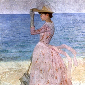
Liverwort spore elaters, light micrograph
Liverwort spore elaters, light micrograph. Transverse section through the sporangium of a liverwort (Pellia epiphylla). This is the basal portion of the sporangium, a region of the sporangium also called the elateraphore. The spores (pink) are mixed up with elongated cells with spiral thickening (elaters). When the sporangium splits open these elaters twist around as they dry and flick out the spores, which are then carried away by air currents. Magnification: x100 when printed at 10 centimetres wide
Science Photo Library features Science and Medical images including photos and illustrations
Media ID 6308393
© DR KEITH WHEELER/SCIENCE PHOTO LIBRARY
Bryophyte Bryophytes Bryophytic Cellular Cross Section Elater Elaters Internal Structure Liverwort Part Parts Plant Anatomy Re Production Reproductive Reproductive Part Reproductive Parts Spore Spores Structural Tissue Transverse Cells Light Micrograph Light Microscope Section Sectioned
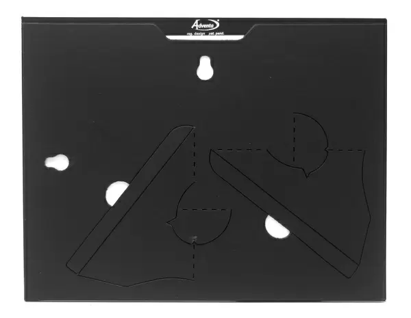
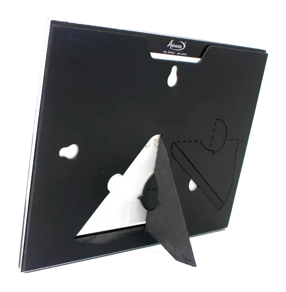
7"x5" Glass Mount
Wall mounted or free-standing, these black edged glass frames feature a smooth chamfered edge and a stylish black border (on back face of the glass). Manufactured from 4mm thick glass, Glass Mounts are a durable, professional way of displaying and protecting your prints. Your 7x5 print is slotted into the back of the frame so can easily be changed if needed.
Tempered Glass Mounts are ideal for wall display, plus the smaller sizes can also be used free-standing via an integral stand
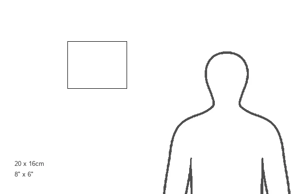
Estimated Image Size (if not cropped) is 17.7cm x 12.7cm (7" x 5")
Estimated Product Size is 20.3cm x 16.2cm (8" x 6.4")
These are individually made so all sizes are approximate
Artwork printed orientated as per the preview above, with landscape (horizontal) orientation to match the source image.
EDITORS COMMENTS
This print showcases the intricate world of liverwort spore elaters, captured under a light microscope. The image reveals a transverse section through the sporangium of a liverwort known as Pellia epiphylla. Specifically, it depicts the basal portion of the sporangium, also referred to as the elateraphore. Within this microcosm, we observe an enthralling interplay between pink spores and elongated cells adorned with spiral thickening called elaters. As nature's ingenious mechanism unfolds, these elaters play a crucial role in dispersing the spores. When the sporangium splits open, these remarkable structures twist around as they dry and suddenly flick out the spores into their surroundings. The mesmerizing visual is not only aesthetically pleasing but also sheds light on an essential aspect of plant reproduction. Magnified at 100 times its original size when printed at 10 centimeters wide, this photograph offers us an intimate glimpse into the internal structure and cellular intricacies of bryophytes like liverworts. By capturing such minute details within botanical anatomy, this image from Science Photo Library invites us to marvel at nature's complexity while deepening our understanding of plant biology. It serves as a testament to both scientific exploration and artistic appreciation for Earth's diverse flora.
MADE IN THE UK
Safe Shipping with 30 Day Money Back Guarantee
FREE PERSONALISATION*
We are proud to offer a range of customisation features including Personalised Captions, Color Filters and Picture Zoom Tools
SECURE PAYMENTS
We happily accept a wide range of payment options so you can pay for the things you need in the way that is most convenient for you
* Options may vary by product and licensing agreement. Zoomed Pictures can be adjusted in the Basket.