
Glass Coaster : Intestinal arteriole, TEM


![]()
Home Decor from Science Photo Library
Intestinal arteriole, TEM
Intestinal arteriole. Transmission electron micrograph (TEM) of a section through an arteriole in the wall of the small intestine. Magnification: x5000 when printed 10 centimetres wide
Science Photo Library features Science and Medical images including photos and illustrations
Media ID 9225753
© STEVE GSCHMEISSNER/SCIENCE PHOTO LIBRARY
Arteriole Basal Lamina Basement Membrane Collagen Elastic Endothelial Endothelium Gastrointestinal System Histological Histology Internal Intestines Layer Lining Membrane Mucosal Muscular Organelle Organelles Outer Small Intestine Smooth Muscle Sub Mucosa Tissue Transmission Electron Micrograph Transmission Electron Microscope Vascular Vesicles Vessels Wall Blood Vessel Cells Circulatory System Section Sectioned
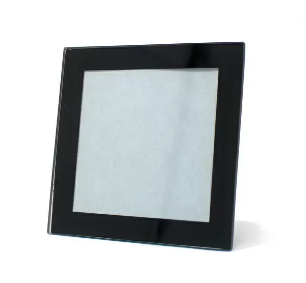
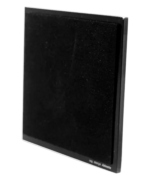
Glass Coaster
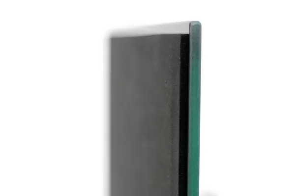
Individual Glass Coaster. Stylish and elegant polished safety glass, toughened and heat resistant (10x10cm, 7mm thick). Price shown is per individual coaster.
Individual Glass Coaster. Elegant polished safety toughened glass and heat resistant, matching Place Mats are also available
Estimated Image Size (if not cropped) is 7.6cm x 6.7cm (3" x 2.6")
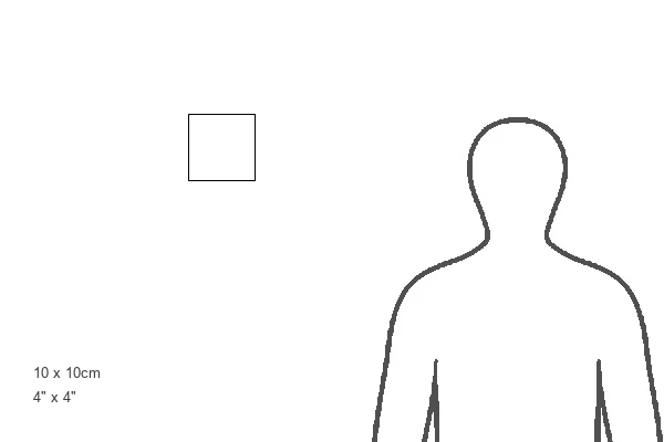
Estimated Product Size is 10cm x 10cm (3.9" x 3.9")
These are individually made so all sizes are approximate
Artwork printed orientated as per the preview above, with landscape (horizontal) orientation to match the source image.
EDITORS COMMENTS
This print from Science Photo Library showcases the intricate details of an intestinal arteriole, providing a glimpse into the complex world of biology and anatomy. Taken using a transmission electron microscope (TEM), this image reveals the inner workings of a blood vessel within the wall of the small intestine. At a magnification of x5000 when printed 10 centimeters wide, every aspect comes to life in stunning clarity. The muscular and collagenous layers can be observed, highlighting their crucial role in maintaining healthy tissue structure. The delicate lining, known as endothelium, is visible along with submucosa and mucosal cells that contribute to proper functioning. The photograph also captures various organelles within these cells, shedding light on their vital functions within the body's circulatory system. From smooth muscle fibers to pinocytotic vesicles involved in cellular uptake processes like pinocytosis, each detail adds depth to our understanding of this biological marvel. With its emphasis on histology and histological structures such as basement membranes and basal laminae, this image serves as a valuable resource for researchers studying gastrointestinal systems or anyone fascinated by human anatomy. It offers both educational value and aesthetic appeal through its meticulous composition. Science Photo Library continues to deliver exceptional visual content that not only educates but also inspires curiosity about the wonders found within our own bodies.
MADE IN THE UK
Safe Shipping with 30 Day Money Back Guarantee
FREE PERSONALISATION*
We are proud to offer a range of customisation features including Personalised Captions, Color Filters and Picture Zoom Tools
SECURE PAYMENTS
We happily accept a wide range of payment options so you can pay for the things you need in the way that is most convenient for you
* Options may vary by product and licensing agreement. Zoomed Pictures can be adjusted in the Basket.