Fraud Blocker Skip to main content

Microscopic Photos Collection (page 3)

Discovering the hidden wonders of the microscopic world is like entering a realm of infinite fascination

Background imageMicroscopic Photos Collection: Human sperm

Human sperm

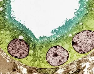
Background imageMicroscopic Photos Collection: Kidney tubule, TEM

Kidney tubule, TEM
Kidney tubule. Coloured transmission electron micrograph (TEM) of a section through a proximal convoluted tubule in the kidney

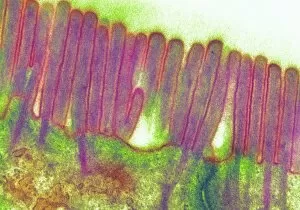
Background imageMicroscopic Photos Collection: Duodenal microvilli

Duodenal microvilli
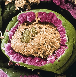
Microvilli in duodenum. Coloured transmission electron micrograph (TEM) of a section through the human duodenum, showing microvilli on the surface epithelium (lining)

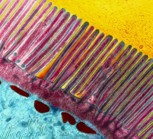
Background imageMicroscopic Photos Collection: Coloured TEM of microvilli of intestinal cell

Coloured TEM of microvilli of intestinal cell

Background imageMicroscopic Photos Collection: Liver capillary, SEM

Liver capillary, SEM
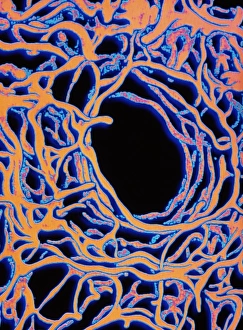
Liver capillary. Coloured scanning electron micro- graph (SEM) of a freeze-fractured section through liver tissue showing a hepatic capillary, or sinusoid

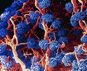
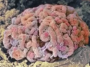
Background imageMicroscopic Photos Collection: Coloured SEM of glomeruli in the human kidney

Coloured SEM of glomeruli in the human kidney

Background imageMicroscopic Photos Collection: Pancreas cell, TEM

Pancreas cell, TEM
Pancreas cell. Coloured transmission electron micrograph (TEM) of an acinar (exocrine) pancreatic cell. Acinar cells secrete the inactive precursors (zymogens)

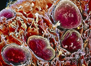
Background imageMicroscopic Photos Collection: Kidney glomerulus

Kidney glomerulus
Glomerulus. Coloured scanning electron micrograph (SEM) of a healthy kidney glomerulus. The glomerulus is a tight ball of capillaries where blood passing through the kidneys is filtered
Background imageMicroscopic Photos Collection: Coloured SEM of a sectioned villus from the ileum

Coloured SEM of a sectioned villus from the ileum

Background imageMicroscopic Photos Collection: Kidney podocyte cells, SEM

Kidney podocyte cells, SEM
Kidney podocyte cells. Coloured scanning electron micrograph (SEM) of podocyte cells on the surface of a kidney glomerulus. Podocytes possess branching, foot-like processes (red/brown)
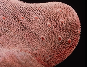
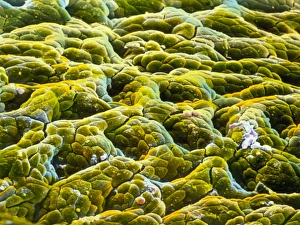
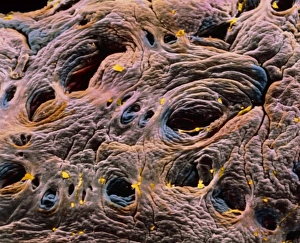
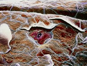
Background imageMicroscopic Photos Collection: False colour SEM of entrance to gastric gland

False colour SEM of entrance to gastric gland
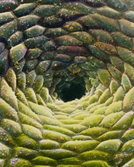
False-colour scanning electron micrograph (SEM) of the mucous membrane lining the stomach, showing the entrance to a gastric gland

Background imageMicroscopic Photos Collection: Stomach wall, SEM

Stomach wall, SEM
Stomach wall. Coloured scanning electron micrograph (SEM) of a section through a freeze- fractured fold in the stomach wall

Background imageMicroscopic Photos Collection: Ear canal, SEM

Ear canal, SEM
Ear canal. Coloured scanning electron micrograph (SEM) of hairs growing in an ear canal. The ear canal runs between the outer ear and the eardrum

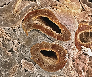
Background imageMicroscopic Photos Collection: Kidney tubules, SEM

Kidney tubules, SEM
Kidney tubules. Coloured scanning electron micrograph (SEM) of proximal convoluted tubules (dark brown) in the kidney. The proximal convoluted tubules function to reabsorb water

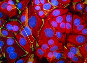
Background imageMicroscopic Photos Collection: Immunofluorescent LM of normal breast cells

Immunofluorescent LM of normal breast cells

Background imageMicroscopic Photos Collection: False-colour SEM of gastric glands of stomach

False-colour SEM of gastric glands of stomach
Gastric gland: false-colour scanning electron micrograph (SEM) of the mucous membrane lining the stomach, showing the shadowy entrances to a number of gastric glands

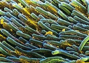
Background imageMicroscopic Photos Collection: Rod cells

Rod cells. Coloured scanning electron micrograph (SEM) of rod cells (green) in the retina of the eye. Rod cells are light-sensitive cells which respond to dim light

Background imageMicroscopic Photos Collection: Purkinje nerve cells

Purkinje nerve cells

Background imageMicroscopic Photos Collection: Coloured SEM of femoral spongy bone

Coloured SEM of femoral spongy bone

Background imageMicroscopic Photos Collection: Bone, SEM

Bone, SEM
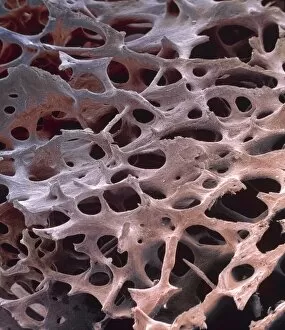
Spongy bone. Coloured scanning electron micrograph (SEM) of freeze-fractured spongy bone tissue. It shows the matrix of collagen and calcium crystals that form the structure of bone tissue

Background imageMicroscopic Photos Collection: Smooth muscle, SEM

Smooth muscle, SEM
Smooth muscle. Coloured scanning electron micrograph of smooth muscle from the trachea (windpipe). Smooth muscle is not under voluntary control, unlike skeletal (striated) muscle

Background imageMicroscopic Photos Collection: Spongy bone, SEM

Spongy bone, SEM
Spongy bone. Coloured scanning electron micrograph (SEM) of human spongy (cancellous) bone from the shaft of a long bone. Found inside bones, below a dense shell of hard compact bone

Background imageMicroscopic Photos Collection: Coloured SEM of section through human spleen

Coloured SEM of section through human spleen
Human spleen. Coloured Scanning Electron Micro- graph (SEM) of a section through the human spleen. Here, the fine network of reticular fibres known as trabeculae is seen

Background imageMicroscopic Photos Collection: Coloured TEM of a basophil white blood cell

Coloured TEM of a basophil white blood cell
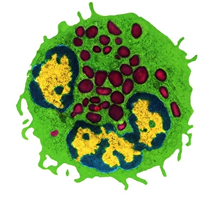
Polynuclear basophil. Coloured Transmission Electron Micrograph (TEM) of a basophil white blood cell. This basophil has one bi-lobed nucleus (yellow, lower centre) and a second nucleus at upper left

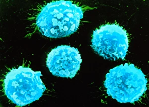
Background imageMicroscopic Photos Collection: False-colour SEM of human T-lymphocytes

False-colour SEM of human T-lymphocytes
False-colour scanning electron micrograph (SEM) of human T-lymphocytes. They are white blood cells, & one of the component cell types of the human immune system

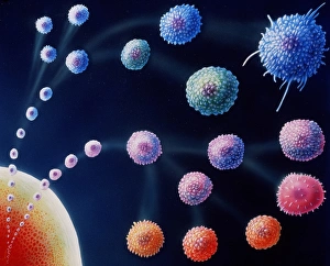
Background imageMicroscopic Photos Collection: White blood cell types

White blood cell types
Artwork featuring different types of white blood cells that comprise the orchestra of the human immune system, the bodys defence mechanism

Background imageMicroscopic Photos Collection: Coloured SEM of B-lymphocyte white blood cells

Coloured SEM of B-lymphocyte white blood cells
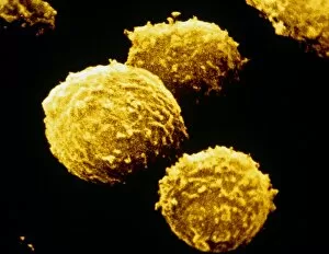
B-lymphocyte. Coloured Scanning Electron Microgr- aph of a group of B-lymphocyte white blood cells. The cell surfaces are covered with tiny ridges and projections called microvilli

Background imageMicroscopic Photos Collection: SEM of lymphocytes in cortex of thymus

SEM of lymphocytes in cortex of thymus
False-colour scanning electron micrograph of the cortex of a thymus. The spheres are T-lymphocytes, white blood cells vital to the cell mediated resp- onse of the immune system

Background imageMicroscopic Photos Collection: TEM of cluster of P1 bacteriophages

TEM of cluster of P1 bacteriophages

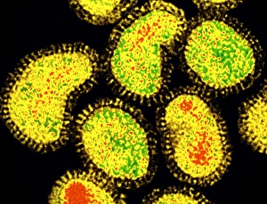
Background imageMicroscopic Photos Collection: Coloured TEM of a cluster of influenza viruses

Coloured TEM of a cluster of influenza viruses

Background imageMicroscopic Photos Collection: Coloured SEM of intestine, showing coeliac disease

Coloured SEM of intestine, showing coeliac disease
Coeliac disease. Coloured Scanning Electron Micro- graph (SEM) of the wall of the small intestine, showing coeliac disease

Background imageMicroscopic Photos Collection: Lacrimal gland, SEM

Lacrimal gland, SEM

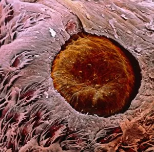
Background imageMicroscopic Photos Collection: SEM of hair on scalp

SEM of hair on scalp
Hair. Coloured scanning electron micrograph of hairs protruding from the surface of the scalp. Numerous desquamating cells (pink) are concentrically arranged around the base of the hair shaft

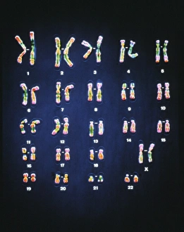
Background imageMicroscopic Photos Collection: Normal female chromosomes

Normal female chromosomes
False-colour light micrograph of a normal trypsin/leishman banded female karyotype, the full complement of female chromosomes arranged in numbered homologous pairs

Background imageMicroscopic Photos Collection: Renal corpuscle of kidney

Renal corpuscle of kidney
Coloured scanning electron micrograph (SEM) of a renal corpuscle in the kidney. The renal corpuscle is formed by the glomerulus (red), the convoluted capillary at centre

Background imageMicroscopic Photos Collection: False-colour SEM of a taste bud on tongue surface

False-colour SEM of a taste bud on tongue surface
Taste bud. False-colour scanning electron micrograph of the apical part of a taste bud (pink at centre), known as a gustatory caliculus, on the dorsal surface of the tongue

Background imageMicroscopic Photos Collection: False-colour SEM of fungiform papillae on tongue

False-colour SEM of fungiform papillae on tongue

Background imageMicroscopic Photos Collection: False-colour SEM of dorsal surface of tongue

False-colour SEM of dorsal surface of tongue
Tongue surface. False-colour scanning electron micrograph (SEM) of the dorsal surface of the tongue. It is covered by two types of projections known as filiform and fungiform papillae

Background imageMicroscopic Photos Collection: False-colour SEM of a papilla vallata

False-colour SEM of a papilla vallata
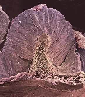
Papilla vallata on tongues surface. False-colour scanning electron micrograph of a papilla vallata (brown), a site of taste buds, partly surrounded by filiform papillae (bottom)


All Professionally Made to Order for Quick Shipping

Discovering the hidden wonders of the microscopic world is like entering a realm of infinite fascination. From particle tracks leaving trails of mystery to the mesmerizing bubble chamber photo capturing the decay of a sigma particle, these microscopic photos unveil secrets that lie beyond our naked eye's reach. One remarkable image showcases the first observation of an omega-minus particle, a groundbreaking moment in scientific history. Meanwhile, another snapshot reveals the delicate beauty of a human blastocyst, reminding us of life's miraculous beginnings. Immunofluorescent LM offers us glimpses into intricate networks within our own bodies - neurons and astrocytes intricately woven together like cosmic constellations. And then there are oxytocin hormone crystals, their vibrant colors shining through as if encapsulating love itself. The iris of an eye captured by SEM reminds us that even something as ordinary as our eyes holds extraordinary complexity when seen up close. Similarly, osteoporotic bone under SEM unveils its fragile structure with astonishing detail. Light micrographs bring forth stunning images such as oxytocin crystals glistening like precious gems or spiral spore chains formed by Streptomyces bacteria resembling ethereal works of art. And let us not forget about our retina - a gateway to vision and perception - or nerve cells firing messages at lightning speed throughout our bodies, orchestrating every movement and thought we have. These microscopic photos offer more than just visual delight; they invite us to ponder upon the vastness contained within each minuscule frame. They remind us that sometimes it is in exploring the smallest details that we gain profound insights into ourselves and the world around us.