Home > Science > SEM
Rod cells. Coloured scanning electron micrograph

![]()
Wall Art and Photo Gifts from Science Photo Library
Rod cells. Coloured scanning electron micrograph
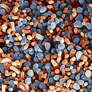
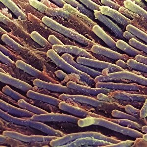
Rod cells. Coloured scanning electron micrograph (SEM) of rod cells (green) in the retina of the eye. Rod cells are light-sensitive cells which respond to dim light, and so are mainly used for dark-adapted vision. They cannot detect colours, which is why vision in low light conditions is limited to shades of grey. Colours are detected by cone cells (not seen). When light falls on the tip of a rod cell, a chemical reaction sends an electrical impulse to the optic nerve (not shown), which relays it to the visual cortex of the brain. Magnification unknown
Science Photo Library features Science and Medical images including photos and illustrations
Media ID 6449077
© STEVE GSCHMEISSNER/SCIENCE PHOTO LIBRARY
Histology Magnified Image Microscopic Photos Retinal Sight Subjects Vision Visual Sense
FEATURES IN THESE COLLECTIONS
EDITORS COMMENTS
This print from Science Photo Library showcases the intricate beauty of rod cells in the retina of the eye. In this coloured scanning electron micrograph (SEM), rod cells are depicted in vibrant green, highlighting their crucial role in our visual perception. Rod cells are specialized light-sensitive receptors that enable us to see in dim lighting conditions. They play a vital role in dark-adapted vision, allowing us to navigate and perceive objects even when there is limited light available. However, these remarkable cells have one limitation - they cannot detect colors. Consequently, our vision during low-light situations is confined to shades of grey. The image also alludes to another type of photoreceptor called cone cells; however, they remain unseen here. Unlike rods, cone cells are responsible for color detection and provide us with a rich palette of hues under normal lighting conditions. When light reaches the tip of a rod cell, it triggers a chemical reaction that generates an electrical impulse. This impulse then travels through the optic nerve (not shown) and ultimately reaches the visual cortex in our brain where it is processed into meaningful images. With its magnification undisclosed but undoubtedly impressive, this microscopic photograph offers a glimpse into the fascinating world within our retinas – an essential component contributing to our sight and overall understanding of human anatomy.
MADE IN THE UK
Safe Shipping with 30 Day Money Back Guarantee
FREE PERSONALISATION*
We are proud to offer a range of customisation features including Personalised Captions, Color Filters and Picture Zoom Tools
SECURE PAYMENTS
We happily accept a wide range of payment options so you can pay for the things you need in the way that is most convenient for you
* Options may vary by product and licensing agreement. Zoomed Pictures can be adjusted in the Basket.