Fraud Blocker Skip to main content

Microscopic Photos Collection (page 2)

Discovering the hidden wonders of the microscopic world is like entering a realm of infinite fascination

Background imageMicroscopic Photos Collection: Blood clot, SEM P260 / 0123

Blood clot, SEM P260 / 0123
Blood clot. Coloured scanning electron micrograph (SEM) of a blood clot. The red blood cells (erythrocytes) are trapped in filaments of fibrin protein (pink)

Background imageMicroscopic Photos Collection: Coloured SEM of the feather of a wren

Coloured SEM of the feather of a wren
Feather. Coloured scanning electron micrograph of barbs, barbules and barbicels on the feather of a wren, Troglodytes troglodytes

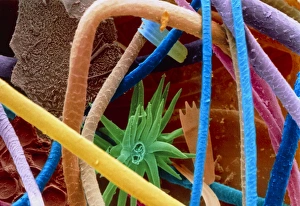
Background imageMicroscopic Photos Collection: Col. SEM of filaments of a tooth brush

Col. SEM of filaments of a tooth brush
Toothbrush. Coloured scanning electron micrograph of the filaments of a toothbrush. Magnification: x37 at 6x7cm size. x74 at 8x6ins, x40 at 9x7cm master size

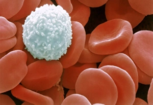
Background imageMicroscopic Photos Collection: Colour SEM of red & white blood cells

Colour SEM of red & white blood cells
Blood cells. Coloured scanning electron micrograph (SEM) of human blood showing red and white cells. Red blood cells (erythrocytes) have a characteristic biconcave-disc shape and are numerous

Background imageMicroscopic Photos Collection: Coloured SEM of household dust (allergens)

Coloured SEM of household dust (allergens)
Household dust. Coloured Scanning Electron Micro- graph (SEM) of a sample of household dust. Dust contains an assortment of substances which can cause asthma or other allergic reactions

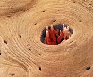
Background imageMicroscopic Photos Collection: Coloured SEM of transverse section of compact bone

Coloured SEM of transverse section of compact bone
Compact bone. Coloured scanning electronmicrograph (SEM) of a transverse section ofcompact bone. The Haversian canal (dark centralarea) contains blood vessels

Background imageMicroscopic Photos Collection: Coloured SEM of the surface of human skin

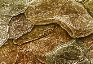
Coloured SEM of the surface of human skin
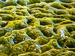
Human skin. Coloured scanning electron micrograph (SEM) of the surface of healthy human skin. The scaly appearance is due to the dead outer cells being constantly shed (sloughed off) and replaced

Background imageMicroscopic Photos Collection: Oxytocin hormone crystals, PLM C016 / 7199

Oxytocin hormone crystals, PLM C016 / 7199
Oxytocin. Polarised light micrograph (PLM) of crystals of the female hormone oxytocin. In women this hormone is secreted naturally by the pituitary gland

Background imageMicroscopic Photos Collection: Oestradiol, female sex hormone, PLM C016 / 7197

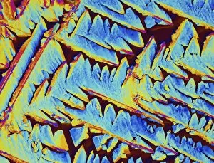
Oestradiol, female sex hormone, PLM C016 / 7197
Female sex hormone. Polarised light micrograph (PLM) of crystals of Oestradiol, also known as beta- Estradiol. This is the most potent of the six naturally-occurring oestrogens

Background imageMicroscopic Photos Collection: Synapse, SEM C018 / 0122

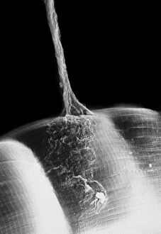
Synapse, SEM C018 / 0122
Synapse. Scanning electron micrograph (SEM) of a neuromuscular junction showing a motor neurone (vertical line) terminating on skeletal muscle fibres (across bottom frame)

Background imageMicroscopic Photos Collection: Synapse, SEM

Synapse, SEM
Synapse. Coloured scanning electron micrograph (SEM) of a neuromuscular junction showing a motor neurone (green) terminating on skeletal muscle fibres (orange)

Background imageMicroscopic Photos Collection: Oestradiol, female sex hormone, PLM C016 / 7213

Oestradiol, female sex hormone, PLM C016 / 7213
Female sex hormone. Polarised light micrograph (PLM) of crystals of Oestradiol, also known as beta- Estradiol. This is the most potent of the six naturally-occurring oestrogens

Background imageMicroscopic Photos Collection: Oestradiol, female sex hormone, PLM C016 / 7210

Oestradiol, female sex hormone, PLM C016 / 7210
Female sex hormone. Polarised light micrograph (PLM) of crystals of Oestradiol, also known as beta- Estradiol. This is the most potent of the six naturally-occurring oestrogens

Background imageMicroscopic Photos Collection: Oestradiol, female sex hormone, PLM C016 / 7202

Oestradiol, female sex hormone, PLM C016 / 7202
Female sex hormone. Polarised light micrograph (PLM) of crystals of Oestradiol, also known as beta- Estradiol. This is the most potent of the six naturally-occurring oestrogens

Background imageMicroscopic Photos Collection: Salt crystal, light micrograph

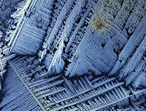
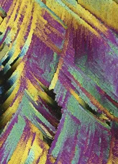
Salt crystal, light micrograph
Salt. Polarised light micrograph of a crystal of sodium chloride, the common salt. This is a white crystalline compound widespread in nature as a solid (rock salt)

Background imageMicroscopic Photos Collection: Testosterone hormone, light micrograph C015 / 6787

Testosterone hormone, light micrograph C015 / 6787
Testosterone hormone. Polarised light micrograph of crystals of the male sex hormone testosterone. Testosterone is the main human androgen hormone

Background imageMicroscopic Photos Collection: Insulin crystals, light micrograph C017 / 8246

Insulin crystals, light micrograph C017 / 8246
Insulin. Polarised light micrograph (PLM) of crystals of the hormone insulin. The insulin molecule is made up of two chains of amino acids (A & B chains)

Background imageMicroscopic Photos Collection: Blue green algae stigonema, LM C013 / 5286

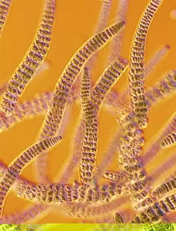
Blue green algae stigonema, LM C013 / 5286
Light micrograph using differential interference contrast microscopy (DIC) of the filamentous blue-green algae (group name Cyanophycophyta), called stigonema

Background imageMicroscopic Photos Collection: SEM of daisy pollen B786 / 0524

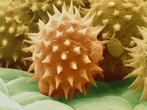
SEM of daisy pollen B786 / 0524
Daisy pollen. Coloured scanning electron micrograph (SEM) of pollen grains of a common Daisy (Bellis perennis). The outer wall (exine) has many small spiky projections

Background imageMicroscopic Photos Collection: Coloured SEM of sunflower pollen grains

Coloured SEM of sunflower pollen grains
Pollen grains. Coloured Scanning Electron Micro- graph of pollen grains of the sunflower, Helianthus annuus. Pollen grain size, shape and surface texture differ from one plant species to another

Background imageMicroscopic Photos Collection: Petal papillae, SEM

Petal papillae, SEM
Petal papillae. Coloured scanning electron micrograph (SEM) of papillae on the surface of a cultivated primrose (Primula obconica) petal. Papillae are projections from epidermal cells

Background imageMicroscopic Photos Collection: Reflective belt material

Reflective belt material
Light micrograph in interference contrast illumination of a reflective material used in safety clothing. The material is retro-reflective

Background imageMicroscopic Photos Collection: Crystals of silver on copper wire

Crystals of silver on copper wire
Light micrograph in incident illumination of crystalline silver deposited on a copper wire. Copper wire is suspended in a solution of silver nitrate

Background imageMicroscopic Photos Collection: LM of melted arsenic disulphide

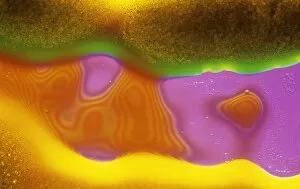
LM of melted arsenic disulphide
Light micrograph of molten arsenic disulphide. Interference contrast. Magnification: x200 at 35mm size

Background imageMicroscopic Photos Collection: Kettle fur limescale

Kettle fur limescale
Kettle fur". Coloured scanning electron micrograph (SEM) of fur from a domestic kettle, showing the crystals that form in a hard water area

Background imageMicroscopic Photos Collection: Eyebrow hair, SEM

Eyebrow hair, SEM
Eyebrow hair and skin. Coloured scanning electron micrograph (SEM) of eyebrow hair growing from the surface of human skin

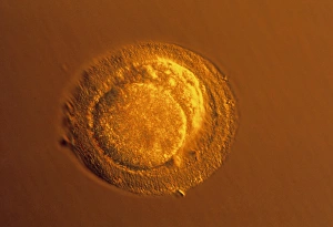
Background imageMicroscopic Photos Collection: LM of four-cell embryo

LM of four-cell embryo
Four-cell embryo. Light micrograph of the blasto- meres of a four-cell human embryo, two days after fertilisation. The blastomeres are the rounded cells

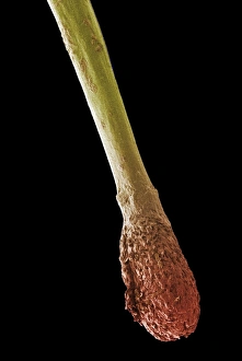
Background imageMicroscopic Photos Collection: Hair root, SEM

Hair root, SEM
Hair root. Coloured scanning electron micrograph (SEM) of the root (red) and part of the shaft (yellow) of a human hair. The base of the hair is known as the hair bulb and is the site of hair growth

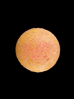
Background imageMicroscopic Photos Collection: Coloured SEM of a fertilized human egg (zygote)

Coloured SEM of a fertilized human egg (zygote)

Background imageMicroscopic Photos Collection: Sperm fertilising egg

Sperm fertilising egg

Background imageMicroscopic Photos Collection: LM of blastomeres of a two-cell embryo

LM of blastomeres of a two-cell embryo
Two-cell embryo. Light micrograph of the blasto- meres of a 2-cell human embryo, two days after fertilisation. The blastomeres are the rounded cells formed by the first division of the fertilized

Background imageMicroscopic Photos Collection: Human sperm

Human sperm

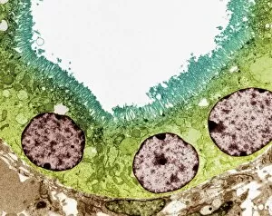
Background imageMicroscopic Photos Collection: Kidney tubule, TEM

Kidney tubule, TEM
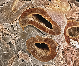
Kidney tubule. Coloured transmission electron micrograph (TEM) of a section through a proximal convoluted tubule in the kidney

Background imageMicroscopic Photos Collection: Duodenal microvilli

Duodenal microvilli
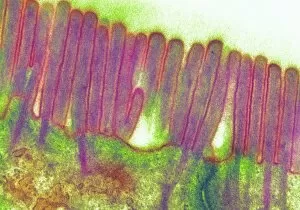
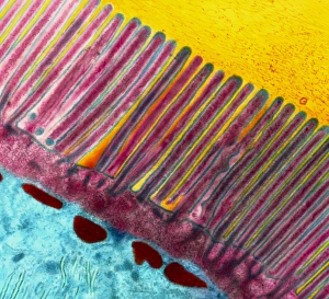
Microvilli in duodenum. Coloured transmission electron micrograph (TEM) of a section through the human duodenum, showing microvilli on the surface epithelium (lining)

Background imageMicroscopic Photos Collection: Coloured TEM of microvilli of intestinal cell

Coloured TEM of microvilli of intestinal cell

Background imageMicroscopic Photos Collection: Liver capillary, SEM

Liver capillary, SEM
Liver capillary. Coloured scanning electron micro- graph (SEM) of a freeze-fractured section through liver tissue showing a hepatic capillary, or sinusoid

Background imageMicroscopic Photos Collection: Coloured SEM of glomeruli in the human kidney

Coloured SEM of glomeruli in the human kidney

Background imageMicroscopic Photos Collection: Pancreas cell, TEM

Pancreas cell, TEM
Pancreas cell. Coloured transmission electron micrograph (TEM) of an acinar (exocrine) pancreatic cell. Acinar cells secrete the inactive precursors (zymogens)

Background imageMicroscopic Photos Collection: Kidney glomerulus

Kidney glomerulus
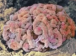
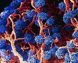
Glomerulus. Coloured scanning electron micrograph (SEM) of a healthy kidney glomerulus. The glomerulus is a tight ball of capillaries where blood passing through the kidneys is filtered
Background imageMicroscopic Photos Collection: Coloured SEM of a sectioned villus from the ileum

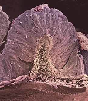
Coloured SEM of a sectioned villus from the ileum

Background imageMicroscopic Photos Collection: Kidney podocyte cells, SEM

Kidney podocyte cells, SEM
Kidney podocyte cells. Coloured scanning electron micrograph (SEM) of podocyte cells on the surface of a kidney glomerulus. Podocytes possess branching, foot-like processes (red/brown)

Background imageMicroscopic Photos Collection: False colour SEM of entrance to gastric gland

False colour SEM of entrance to gastric gland
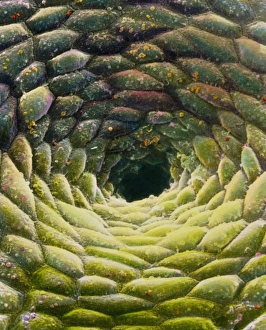
False-colour scanning electron micrograph (SEM) of the mucous membrane lining the stomach, showing the entrance to a gastric gland

Background imageMicroscopic Photos Collection: Stomach wall, SEM

Stomach wall, SEM
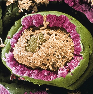
Stomach wall. Coloured scanning electron micrograph (SEM) of a section through a freeze- fractured fold in the stomach wall

Background imageMicroscopic Photos Collection: Ear canal, SEM

Ear canal, SEM
Ear canal. Coloured scanning electron micrograph (SEM) of hairs growing in an ear canal. The ear canal runs between the outer ear and the eardrum

Background imageMicroscopic Photos Collection: Kidney tubules, SEM

Kidney tubules, SEM
Kidney tubules. Coloured scanning electron micrograph (SEM) of proximal convoluted tubules (dark brown) in the kidney. The proximal convoluted tubules function to reabsorb water

Background imageMicroscopic Photos Collection: Immunofluorescent LM of normal breast cells

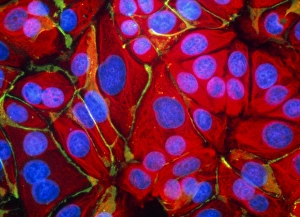
Immunofluorescent LM of normal breast cells

Background imageMicroscopic Photos Collection: False-colour SEM of gastric glands of stomach

False-colour SEM of gastric glands of stomach
Gastric gland: false-colour scanning electron micrograph (SEM) of the mucous membrane lining the stomach, showing the shadowy entrances to a number of gastric glands

Background imageMicroscopic Photos Collection: Rod cells

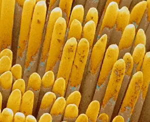
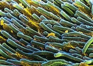
Rod cells. Coloured scanning electron micrograph (SEM) of rod cells (green) in the retina of the eye. Rod cells are light-sensitive cells which respond to dim light


All Professionally Made to Order for Quick Shipping

Discovering the hidden wonders of the microscopic world is like entering a realm of infinite fascination. From particle tracks leaving trails of mystery to the mesmerizing bubble chamber photo capturing the decay of a sigma particle, these microscopic photos unveil secrets that lie beyond our naked eye's reach. One remarkable image showcases the first observation of an omega-minus particle, a groundbreaking moment in scientific history. Meanwhile, another snapshot reveals the delicate beauty of a human blastocyst, reminding us of life's miraculous beginnings. Immunofluorescent LM offers us glimpses into intricate networks within our own bodies - neurons and astrocytes intricately woven together like cosmic constellations. And then there are oxytocin hormone crystals, their vibrant colors shining through as if encapsulating love itself. The iris of an eye captured by SEM reminds us that even something as ordinary as our eyes holds extraordinary complexity when seen up close. Similarly, osteoporotic bone under SEM unveils its fragile structure with astonishing detail. Light micrographs bring forth stunning images such as oxytocin crystals glistening like precious gems or spiral spore chains formed by Streptomyces bacteria resembling ethereal works of art. And let us not forget about our retina - a gateway to vision and perception - or nerve cells firing messages at lightning speed throughout our bodies, orchestrating every movement and thought we have. These microscopic photos offer more than just visual delight; they invite us to ponder upon the vastness contained within each minuscule frame. They remind us that sometimes it is in exploring the smallest details that we gain profound insights into ourselves and the world around us.